• 文献检索
  • 文档翻译
  • 深度研究
  • 学术资讯
  • Suppr Zotero 插件Zotero 插件
  • 邀请有礼
  • 套餐&价格
  • 历史记录
应用&插件
Suppr Zotero 插件Zotero 插件浏览器插件Mac 客户端Windows 客户端微信小程序
定价
高级版会员购买积分包购买API积分包
服务
文献检索文档翻译深度研究API 文档MCP 服务
关于我们
关于 Suppr公司介绍联系我们用户协议隐私条款
关注我们

Suppr 超能文献

核心技术专利:CN118964589B侵权必究
粤ICP备2023148730 号-1Suppr @ 2026

文献检索

告别复杂PubMed语法,用中文像聊天一样搜索,搜遍4000万医学文献。AI智能推荐,让科研检索更轻松。

立即免费搜索

文件翻译

保留排版,准确专业,支持PDF/Word/PPT等文件格式,支持 12+语言互译。

免费翻译文档

深度研究

AI帮你快速写综述,25分钟生成高质量综述,智能提取关键信息,辅助科研写作。

立即免费体验

相似文献

1
Spatiotemporal features of neurogenesis in the retina of medaka, Oryzias latipes.青鳉(Oryzias latipes)视网膜神经发生的时空特征。
Dev Dyn. 2008 Dec;237(12):3870-81. doi: 10.1002/dvdy.21797.
2
Igf signaling couples retina growth with body growth by modulating progenitor cell division.IGF 信号通过调节祖细胞分裂将视网膜生长与身体生长联系起来。
Development. 2021 Apr 1;148(7). doi: 10.1242/dev.199133. Epub 2021 Apr 15.
3
[Development of medaka embryo under simulated microgravity by 3D-clinostat: development of retina and cartilage].[利用三维回转器模拟微重力条件下青鳉胚胎的发育:视网膜和软骨的发育]
Biol Sci Space. 1999 Sep;13(3):162-3.
4
Expression of membrane-bound and soluble guanylyl cyclase mRNAs in embryonic and adult retina of the medaka fish Oryzias latipes.青鳉(Oryzias latipes)胚胎和成年视网膜中膜结合型和可溶性鸟苷酸环化酶mRNA的表达
Zoolog Sci. 2003 Feb;20(2):133-40. doi: 10.2108/zsj.20.133.
5
Genomic organization and embryonic expression of miR-430 in medaka (Oryzias latipes): insights into the post-transcriptional gene regulation in early development.青鳉(Oryzias latipes)中miR-430的基因组组织与胚胎表达:对早期发育中转录后基因调控的见解
Gene. 2010 Jan 1;449(1-2):41-9. doi: 10.1016/j.gene.2009.09.005. Epub 2009 Sep 19.
6
Comparative analysis of her genes during fish somitogenesis suggests a mouse/chick-like mode of oscillation in medaka.在鱼类体节发生过程中对其基因进行的比较分析表明,青鳉中存在类似小鼠/鸡的振荡模式。
Dev Genes Evol. 2006 Jun;216(6):315-32. doi: 10.1007/s00427-006-0059-6. Epub 2006 Mar 17.
7
Evolutionary relationships and diversification of barhl genes within retinal cell lineages.视网膜细胞谱系中 Barhl 基因的进化关系和多样化。
BMC Evol Biol. 2011 Nov 21;11:340. doi: 10.1186/1471-2148-11-340.
8
Genetic developmental timing revealed by inter-species transplantations in fish.鱼类种间移植揭示的遗传发育时序。
Development. 2020 Nov 23;147(22):dev192500. doi: 10.1242/dev.192500.
9
her7 and hey1, but not lunatic fringe show dynamic expression during somitogenesis in medaka (Oryzias latipes).在青鳉(日本青鳉)的体节发生过程中,her7和hey1呈现动态表达,但lunatic fringe没有。
Gene Expr Patterns. 2004 Sep;4(5):553-9. doi: 10.1016/j.modgep.2004.02.003.
10
Specification of primordial germ cells in medaka (Oryzias latipes).青鳉(日本青鳉)中原始生殖细胞的特化
BMC Dev Biol. 2007 Jan 11;7:3. doi: 10.1186/1471-213X-7-3.

引用本文的文献

1
Genetic analysis of medaka fish illuminates conserved and divergent roles of Pax6 in vertebrate eye development.青鳉鱼的遗传分析揭示了Pax6在脊椎动物眼睛发育中的保守和不同作用。
Front Cell Dev Biol. 2024 Oct 24;12:1448773. doi: 10.3389/fcell.2024.1448773. eCollection 2024.
2
Knockout of and in Medaka () Reveals Their Roles in Regulating Vision-Guided Behavior and Eye Development.Medaka 中 和 的敲除揭示了它们在调节视觉引导行为和眼睛发育中的作用。
Int J Mol Sci. 2023 May 15;24(10):8786. doi: 10.3390/ijms24108786.
3
Retinal histogenesis in an altricial avian species, the zebra finch (Taeniopygia guttata, Vieillot 1817).晚成鸟斑马雀(Taeniopygia guttata,Vieillot 1817)的视网膜发生。
J Anat. 2018 Jul;233(1):106-120. doi: 10.1111/joa.12809. Epub 2018 Mar 26.
4
Cell and small animal models for phenotypic drug discovery.用于表型药物发现的细胞和小动物模型。
Drug Des Devel Ther. 2017 Jun 28;11:1957-1967. doi: 10.2147/DDDT.S129447. eCollection 2017.
5
TGF-β Controls miR-181/ERK Regulatory Network during Retinal Axon Specification and Growth.转化生长因子-β在视网膜轴突特化和生长过程中调控miR-181/细胞外信号调节激酶调控网络。
PLoS One. 2015 Dec 7;10(12):e0144129. doi: 10.1371/journal.pone.0144129. eCollection 2015.
6
Workshop report: The medaka model for comparative assessment of human disease mechanisms.研讨会报告:用于人类疾病机制比较评估的青鳉模型
Comp Biochem Physiol C Toxicol Pharmacol. 2015 Dec;178:156-162. doi: 10.1016/j.cbpc.2015.06.003. Epub 2015 Jun 19.
7
Differential responsiveness of distinct retinal domains to Atoh7.不同视网膜区域对Atoh7的差异反应性。
Mech Dev. 2014 Aug;133:218-29. doi: 10.1016/j.mod.2014.08.002. Epub 2014 Aug 21.
8
Tetrahydroquinoline-derived macrocyclic toolbox: the discovery of antiangiogenesis agents in zebrafish assay.四氢喹啉衍生的大环工具库:斑马鱼实验中抗血管生成剂的发现
ACS Med Chem Lett. 2013 May 24;4(7):666-70. doi: 10.1021/ml400026n. eCollection 2013 Jul 11.
9
Chronotopographical distribution patterns of cell death and of lectin-positive macrophages/microglial cells during the visual system ontogeny of the small-spotted catshark Scyliorhinus canicula.小星鲨 Scyliorhinus canicula 视觉系统发生过程中细胞死亡和凝集素阳性巨噬细胞/小胶质细胞的时空分布模式。
J Anat. 2013 Aug;223(2):171-84. doi: 10.1111/joa.12071. Epub 2013 Jun 13.
10
Crb apical polarity proteins maintain zebrafish retinal cone mosaics via intercellular binding of their extracellular domains.Crb 顶端极性蛋白通过其细胞外结构域的细胞间结合维持斑马鱼视网膜锥体镶嵌。
Dev Cell. 2012 Jun 12;22(6):1261-74. doi: 10.1016/j.devcel.2012.03.007. Epub 2012 May 10.

本文引用的文献

1
In vivo validation of a computationally predicted conserved Ath5 target gene set.计算预测的保守Ath5靶基因集的体内验证
PLoS Genet. 2007 Sep;3(9):1661-71. doi: 10.1371/journal.pgen.0030159. Epub 2007 Aug 1.
2
Mechanism of positioning the cell nucleus in vertebrate photoreceptors.脊椎动物光感受器中细胞核定位的机制。
Proc Natl Acad Sci U S A. 2007 Sep 11;104(37):14819-24. doi: 10.1073/pnas.0700178104. Epub 2007 Sep 4.
3
SARA-regulated vesicular targeting underlies formation of the light-sensing organelle in mammalian rods.SARA调节的囊泡靶向作用是哺乳动物视杆细胞中光感受细胞器形成的基础。
Cell. 2007 Aug 10;130(3):535-47. doi: 10.1016/j.cell.2007.06.030.
4
The ultraviolet opsin is the first opsin expressed during retinal development of salmonid fishes.紫外视蛋白是鲑科鱼类视网膜发育过程中表达的首个视蛋白。
Invest Ophthalmol Vis Sci. 2007 Feb;48(2):866-73. doi: 10.1167/iovs.06-0442.
5
The zebrafish genome in context: ohnologs gone missing.斑马鱼基因组的背景情况:同源多倍体基因的缺失
J Exp Zool B Mol Dev Evol. 2007 Sep 15;308(5):563-77. doi: 10.1002/jez.b.21137.
6
Individual cell migration serves as the driving force for optic vesicle evagination.单个细胞迁移是视泡外翻的驱动力。
Science. 2006 Aug 25;313(5790):1130-4. doi: 10.1126/science.1127144.
7
Functional characterization of visual opsin repertoire in Medaka (Oryzias latipes).青鳉(Oryzias latipes)视觉视蛋白库的功能特征
Gene. 2006 Apr 26;371(2):268-78. doi: 10.1016/j.gene.2005.12.005. Epub 2006 Feb 7.
8
Photoreceptor layer of salmonid fishes: transformation and loss of single cones in juvenile fish.鲑科鱼类的光感受器层:幼鱼中单锥体的转变与丧失
J Comp Neurol. 2006 Mar 10;495(2):213-35. doi: 10.1002/cne.20879.
9
Forward genetic analysis of visual behavior in zebrafish.斑马鱼视觉行为的正向遗传学分析。
PLoS Genet. 2005 Nov;1(5):e66. doi: 10.1371/journal.pgen.0010066. Epub 2005 Nov 25.
10
Mammalian inscuteable regulates spindle orientation and cell fate in the developing retina.哺乳动物的无柄蛋白在发育中的视网膜中调节纺锤体方向和细胞命运。
Neuron. 2005 Nov 23;48(4):539-45. doi: 10.1016/j.neuron.2005.09.030.

青鳉(Oryzias latipes)视网膜神经发生的时空特征。

Spatiotemporal features of neurogenesis in the retina of medaka, Oryzias latipes.

作者信息

Kitambi Satish S, Malicki Jarema J

机构信息

School of Life Sciences, Södertörns University College, Stockholm, Sweden.

出版信息

Dev Dyn. 2008 Dec;237(12):3870-81. doi: 10.1002/dvdy.21797.

DOI:10.1002/dvdy.21797
PMID:19035349
原文链接:https://pmc.ncbi.nlm.nih.gov/articles/PMC2919814/
Abstract

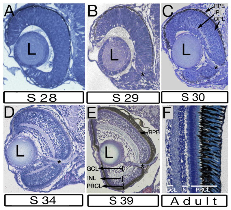
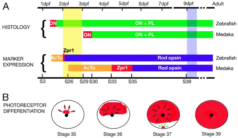
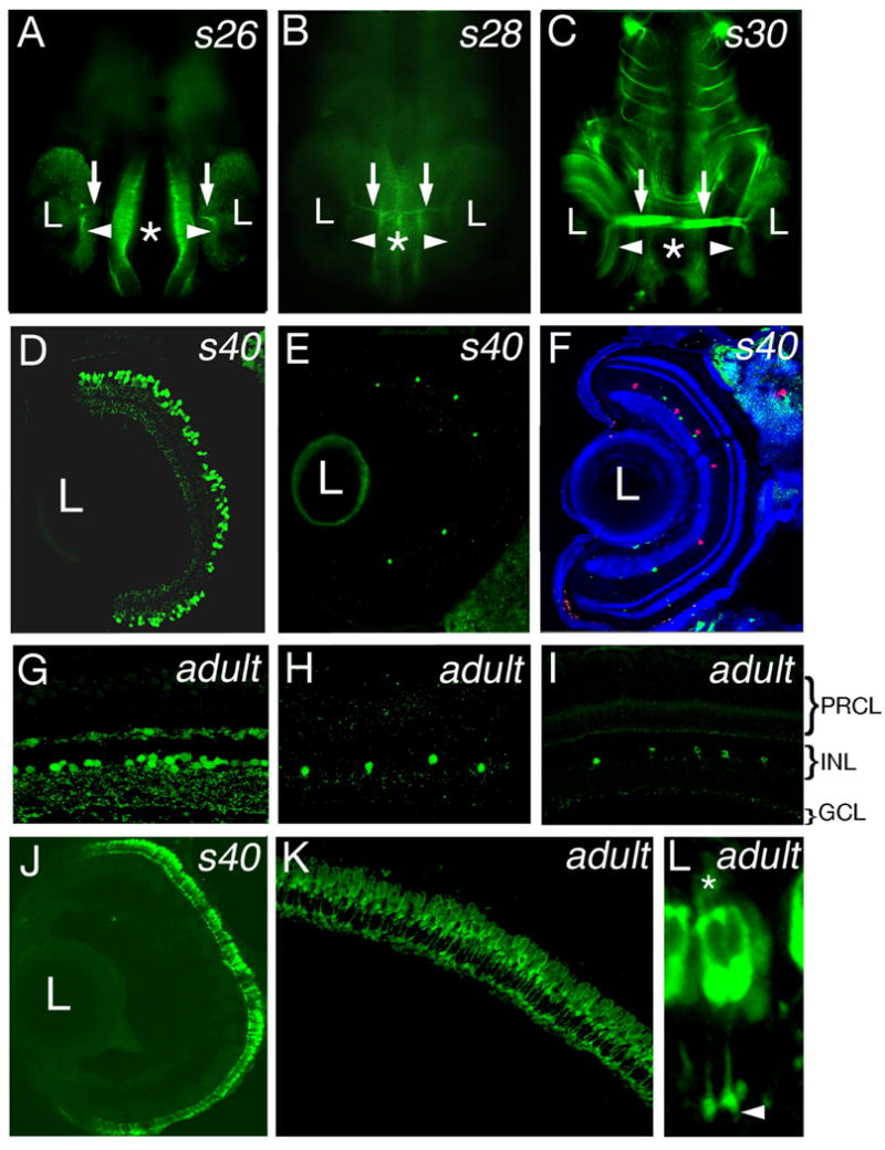
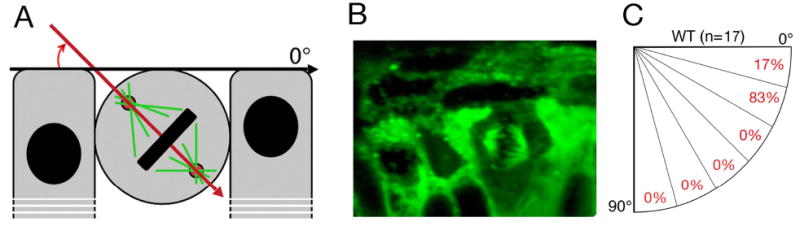
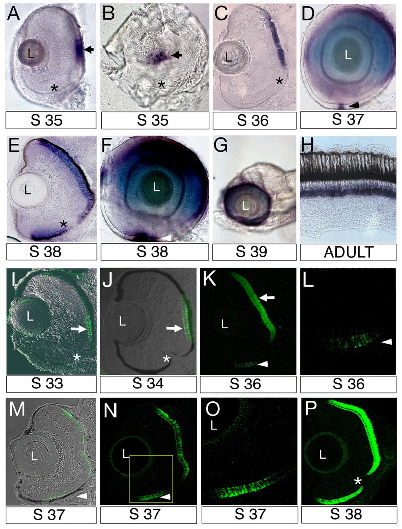
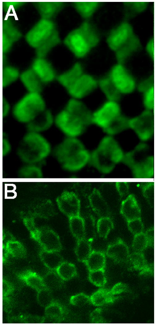
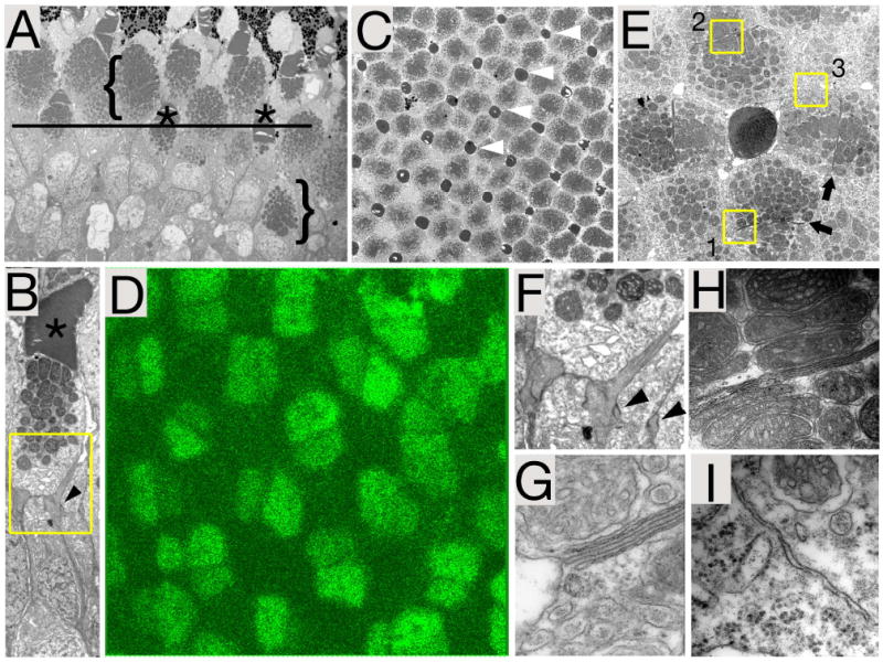
The vertebrate retina is very well conserved in evolution. Its structure and functional features are very similar in phyla as different as primates and teleost fish. Here, we describe the spatiotemporal characteristics of neurogenesis in the retina of a teleost, medaka, and compare them with other species, primarily the zebrafish. Several intriguing differences are observed between medaka and zebrafish. For example, photoreceptor differentiation in the medaka retina starts independently in two different areas, and at more advanced stages of differentiation, medaka and zebrafish retinae display obviously different patterns of the photoreceptor cell mosaic. Medaka and zebrafish evolutionary lineages are thought to have separated from each other 110 million years ago, and so the differences between these species are not unexpected, and may be exploited to gain insight into the architecture of developmental pathways. Importantly, this work highlights the benefits of using multiple teleost models in parallel to understand a developmental process.

摘要

脊椎动物的视网膜在进化过程中得到了很好的保留。其结构和功能特征在灵长类动物和硬骨鱼等不同门类中非常相似。在这里,我们描述了硬骨鱼青鳉视网膜神经发生的时空特征,并将它们与其他物种(主要是斑马鱼)进行比较。在青鳉和斑马鱼之间观察到了几个有趣的差异。例如,青鳉视网膜中的光感受器分化在两个不同区域独立开始,并且在分化的更高级阶段,青鳉和斑马鱼的视网膜显示出明显不同的光感受器细胞镶嵌模式。青鳉和斑马鱼的进化谱系被认为在1.1亿年前彼此分离,因此这些物种之间的差异并不意外,并且可以利用这些差异来深入了解发育途径的结构。重要的是,这项工作强调了并行使用多种硬骨鱼模型来理解发育过程的好处。

https://cdn.ncbi.nlm.nih.gov/pmc/blobs/46cd/2919814/a709e4b59a24/nihms191845f7.jpg
https://cdn.ncbi.nlm.nih.gov/pmc/blobs/46cd/2919814/1e6704f783ee/nihms191845f1.jpg
https://cdn.ncbi.nlm.nih.gov/pmc/blobs/46cd/2919814/2434f4fa1442/nihms191845f2.jpg
https://cdn.ncbi.nlm.nih.gov/pmc/blobs/46cd/2919814/6f73e454f09e/nihms191845f3.jpg
https://cdn.ncbi.nlm.nih.gov/pmc/blobs/46cd/2919814/f47582a30974/nihms191845f4.jpg
https://cdn.ncbi.nlm.nih.gov/pmc/blobs/46cd/2919814/546a5e739d6c/nihms191845f5.jpg
https://cdn.ncbi.nlm.nih.gov/pmc/blobs/46cd/2919814/f6da04a80beb/nihms191845f6.jpg
https://cdn.ncbi.nlm.nih.gov/pmc/blobs/46cd/2919814/a709e4b59a24/nihms191845f7.jpg