Teo Karen S L, Dundon Benjamin K, Molaee Payman, Williams Kerry F, Carbone Angelo, Brown Michael A, Worthley Matthew I, Disney Patrick J, Sanders Prashanthan, Worthley Stephen G
Cardiovascular Research Centre, Royal Adelaide Hospital and The University of Adelaide, Adelaide, South Australia, Australia.
J Cardiovasc Magn Reson. 2008 Dec 1;10(1):55. doi: 10.1186/1532-429X-10-55.
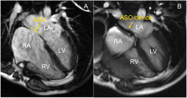
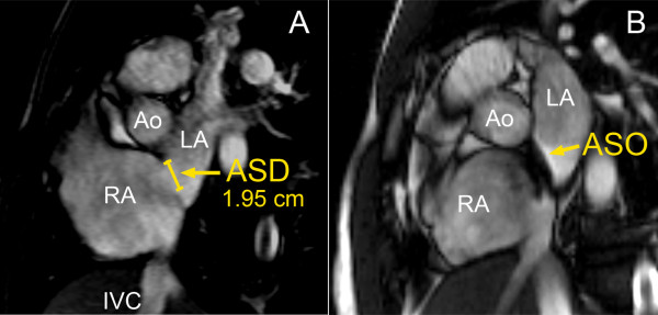
Percutaneous closure of atrial septal defects (ASDs) should potentially reduce right heart volumes by removing left-to-right shunting. Due to ventricular interdependence, this may be associated with impaired left ventricular filling and potentially function. Furthermore, atrial changes post-ASD closure have been poorly understood and may be important for understanding risk of atrial arrhythmia post-ASD closure. Cardiovascular magnetic resonance (CMR) is an accurate and reproducible imaging modality for the assessment of cardiac function and volumes. We assessed cardiac volumes pre- and post-percutaneous ASD closure using CMR.
Consecutive patients (n = 23) underwent CMR pre- and 6 months post-ASD closure. Steady state free precession cine CMR was performed using contiguous slices in both short and long axis views through the ASD. Data was collected for assessment of left and right atrial, ventricular end diastolic volumes (EDV) and end systolic volumes (ESV). Data is presented as mean +/- SD, volumes as mL, and paired t-testing performed between groups. Statistical significance was taken as p < 0.05.
There was a significant reduction in right ventricular volumes at 6 months post-ASD closure (RVEDV: 208.7 +/- 76.7 vs. 140.6 +/- 60.4 mL, p < 0.0001) and RVEF was significantly increased (RVEF 35.5 +/- 15.5 vs. 42.0 +/- 15.2%, p = 0.025). There was a significant increase in the left ventricular volumes (LVEDV 84.8 +/- 32.3 vs. 106.3 +/- 38.1 mL, p = 0.003 and LVESV 37.4 +/- 20.9 vs. 46.8 +/- 18.5 mL, p = 0.016). However, there was no significant difference in LVEF and LV mass post-ASD closure. There was a significant reduction in right atrial volumes at 6 months post-ASD closure (pre-closure 110.5 +/- 55.7 vs. post-closure 90.7 +/- 69.3 mL, p = 0.019). Although there was a trend to a decrease in left atrial volumes post-ASD closure, this was not statistically significant (84.5 +/- 34.8 mL to 81.8 +/- 44.2 mL, p = NS).
ASD closure leads to normalisation of ventricular volumes and also a reduction in right atrial volume. Further follow-up is required to assess how this predicts outcomes such as risk of atrial arrhythmias after such procedures.
经皮闭合房间隔缺损(ASD)可能通过消除左向右分流来减少右心容量。由于心室相互依存关系,这可能与左心室充盈受损及潜在功能障碍有关。此外,ASD闭合术后心房的变化尚未得到充分了解,这对于理解ASD闭合术后房性心律失常的风险可能很重要。心血管磁共振成像(CMR)是一种准确且可重复的成像方式,用于评估心脏功能和容量。我们使用CMR评估了经皮ASD闭合术前和术后的心脏容量。
连续23例患者在ASD闭合术前和术后6个月接受CMR检查。通过ASD在短轴和长轴视图中使用连续切片进行稳态自由进动电影CMR检查。收集数据以评估左、右心房和心室的舒张末期容积(EDV)和收缩末期容积(ESV)。数据以平均值±标准差表示,容积单位为mL,并在组间进行配对t检验。统计学显著性以p<0.05为准。
ASD闭合术后6个月,右心室容量显著减少(RVEDV:208.7±76.7 vs. 140.6±60.4 mL,p<0.0001),右心室射血分数(RVEF)显著增加(RVEF 35.5±15.5 vs. 42.0±15.2%,p = 0.025)。左心室容量显著增加(LVEDV 84.8±32.3 vs. 106.3±38.1 mL,p = 0.003;LVESV 37.4±20.9 vs. 46.8±18.5 mL,p = 0.016)。然而,ASD闭合术后左心室射血分数和左心室质量无显著差异。ASD闭合术后6个月,右心房容量显著减少(术前110.5±55.7 vs. 术后90.7±69.3 mL,p = 0.019)。虽然ASD闭合术后左心房容量有下降趋势,但差异无统计学意义(84.5±34.8 mL至81.8±44.2 mL,p = 无统计学意义)。
ASD闭合可使心室容量正常化,并减少右心房容量。需要进一步随访以评估这如何预测此类手术后的房性心律失常风险等结果。