Radiology, Brigham and Women's Hospital/Harvard Medical School, Boston, MA 02115, USA.
Osteoarthritis Cartilage. 2009 Jun;17(6):761-5. doi: 10.1016/j.joca.2008.11.001. Epub 2008 Nov 12.
To establish the performance of location specific computer measures of radiographic joint space width (JSW) compared to measurements of minimum joint space width (mJSW) for the assessment of medial compartment knee osteoarthritis (OA). The study also investigated the most disease-responsive location for measuring medial compartment JSW.
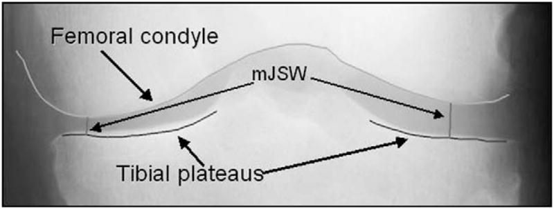
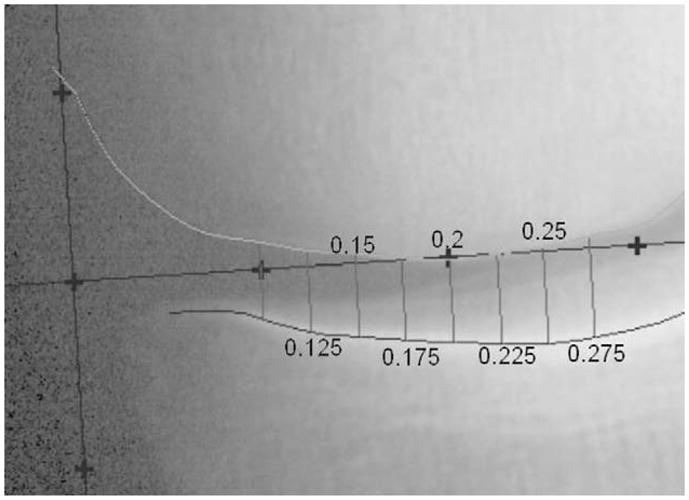
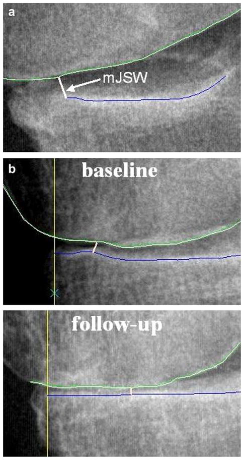
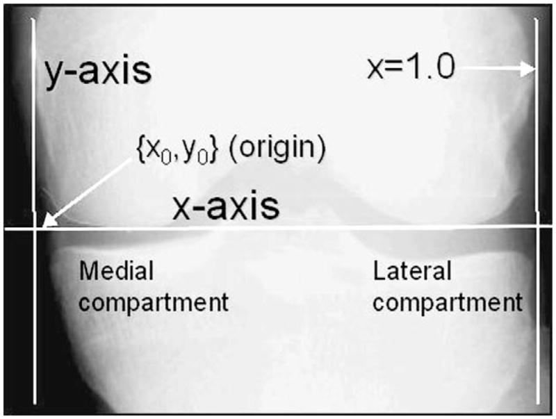
Serial bilateral Posterior Anterior (PA) conventional radiographs acquired with a fixed flexion protocol were obtained 36 months apart in 118 persons with knee OA participating in the Health, Aging and Body Composition (Health ABC) Study. Measurements of medial compartment mJSW and JSW at seven fixed locations were facilitated by the use of semi-automated software that delineated the femoral and tibial margins of the joint. A human reader operated custom software to verify and correct the software-drawn margins where necessary. Paired images were displayed with the reader blinded to the chronological order. The amount of joint space narrowing was measured and the standardized response mean (SRM) was used as a metric to quantify performance.
For all subjects, the mJSW SRM value was 0.42 while, for the most responsive location specific measure of JSW, it was SRM=0.46. For subjects with a Kellgren-Lawrence (KL) score less than or equal to 1, mJSW (SRM=0.40) was more responsive than the new measures (Maximum SRM=0.30). For KL=2or3, SRM=0.49 for mJSW, and SRM=0.74 for the most responsive location specific measure of JSW. Improved responsiveness was observed in the more central portion of the joint on the more diseased knees.
Location specific computer measures of JSW are feasible and potentially provide a superior method to assess radiographic OA for more diseased subjects. This new measure has the potential to improve the power of clinical studies that use a fixed flexion protocol.
建立特定部位计算机测量放射学关节间隙宽度(JSW)与最小关节间隙宽度(mJSW)测量在评估内侧间室膝骨关节炎(OA)中的表现。本研究还探讨了测量内侧间室 JSW 最具疾病反应性的部位。
118 名膝骨关节炎患者参与了健康、衰老和身体成分研究(Health ABC),在 36 个月的时间内,以固定屈曲方案获得了双侧后前(PA)常规放射照片。使用半自动软件方便地测量了内侧间室 mJSW 和七个固定位置的 JSW,该软件勾勒出关节的股骨和胫骨边缘。人工读者使用定制软件来验证和纠正必要的软件绘制的边缘。显示配对图像时,读者对时间顺序是盲目的。测量关节间隙狭窄的程度,并使用标准化反应均值(SRM)作为量化性能的指标。
对于所有受试者,mJSW 的 SRM 值为 0.42,而对于最敏感的特定部位 JSW 测量值,其 SRM 值为 0.46。对于 KL 评分小于或等于 1 的受试者,mJSW(SRM=0.40)比新测量值更敏感(最大 SRM=0.30)。对于 KL=2 或 3,mJSW 的 SRM=0.49,最敏感的特定部位 JSW 测量值的 SRM=0.74。在膝关节病变较重的关节中心部位观察到了更好的反应性。
特定部位的 JSW 计算机测量是可行的,并且可能为更严重的患者提供评估放射学 OA 的更好方法。这种新的测量方法有可能提高使用固定屈曲方案的临床研究的效能。