Department of Brain & Cognitive Sciences, Massachusetts Institute of Technology 46-5121, Cambridge, MA 02139, United States.
Neurobiol Aging. 2010 Nov;31(11):1912-26. doi: 10.1016/j.neurobiolaging.2008.10.015. Epub 2008 Dec 16.
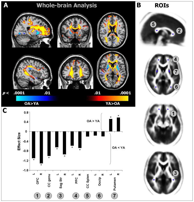
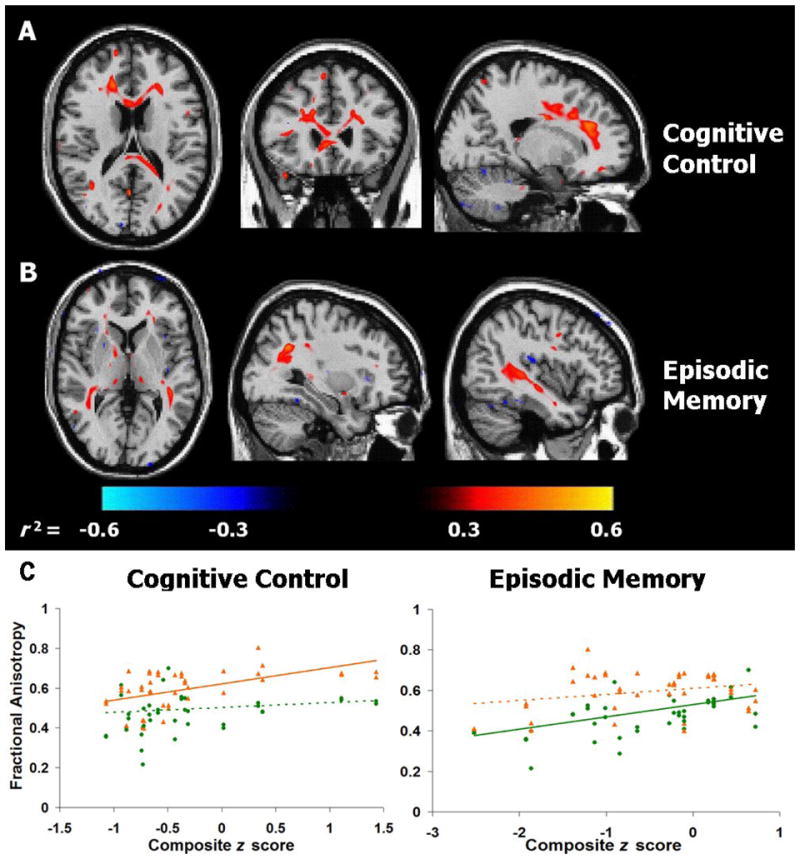
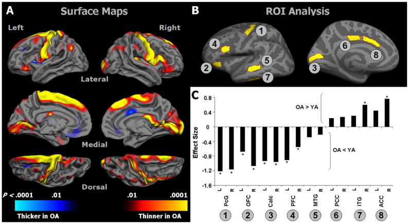
It is well established that healthy aging is accompanied by structural changes in many brain regions and functional decline in a number of cognitive domains. The goal of this study was to determine (1) whether the regional distribution of age-related brain changes is similar in gray matter (GM) and white matter (WM) regions, or whether these two tissue types are affected differently by aging, and (2) whether measures of cognitive performance are more closely linked to alterations in the cerebral cortex or in the underlying WM in older adults (OA). To address these questions, we collected high-resolution magnetic resonance imaging (MRI) data from a large sample of healthy young adults (YA; aged 18-28) and OA (aged 61-86 years). In addition, the OA completed a series of tasks selected to assess cognition in three domains: cognitive control, episodic memory, and semantic memory. Using advanced techniques for measuring cortical thickness and WM integrity, we found that healthy aging was accompanied by deterioration of both GM and WM, but with distinct patterns of change: Cortical thinning occurred primarily in primary sensory and motor cortices, whereas WM changes were localized to regions underlying association cortices. Further, in OA, we found a striking pattern of region-specific correlations between measures of cognitive performance and WM integrity, but not cortical thickness. Specifically, cognitive control correlated with integrity of frontal lobe WM, whereas episodic memory was related to integrity of temporal and parietal lobe WM. Thus, age-related impairments in specific cognitive capacities may arise from degenerative processes that affect the underlying connections of their respective neural networks.
众所周知,健康的衰老伴随着许多大脑区域的结构变化和许多认知领域的功能下降。本研究的目的是确定:(1) 年龄相关的大脑变化在灰质 (GM) 和白质 (WM) 区域的分布是否相似,或者这两种组织类型是否受到衰老的不同影响,以及 (2) 认知表现的衡量标准是否与老年人大脑皮层或下 WM 的变化更密切相关。为了解决这些问题,我们从大量健康的年轻成年人 (YA;年龄 18-28 岁) 和老年人 (OA;年龄 61-86 岁) 中收集了高分辨率磁共振成像 (MRI) 数据。此外,OA 完成了一系列任务,这些任务旨在评估认知的三个领域:认知控制、情景记忆和语义记忆。我们使用测量皮质厚度和 WM 完整性的先进技术,发现健康的衰老伴随着 GM 和 WM 的恶化,但变化模式不同:皮质变薄主要发生在初级感觉和运动皮层,而 WM 变化局限于联合皮层下区域。此外,我们发现 OA 中认知表现与 WM 完整性之间存在惊人的区域特异性相关性,而与皮质厚度无关。具体来说,认知控制与额叶 WM 的完整性相关,而情景记忆与颞叶和顶叶 WM 的完整性相关。因此,特定认知能力的年龄相关损伤可能源于影响其各自神经网络的基础连接的退行性过程。