Erbay S, Han R, Aftab M, Zou Kelly H, Polak J F, Bhadelia Rafeeque A
Department of Radiology, Tufts-New England Medical Center, Boston, MA, USA.
BMC Neurol. 2008 Dec 22;8:51. doi: 10.1186/1471-2377-8-51.
Our purpose was to study the association between the intracranial atherosclerosis as measured by cavernous carotid artery calcification (ICAC) observed on head CT and atrophic changes of supra-tentorial brain demonstrated by MRI.
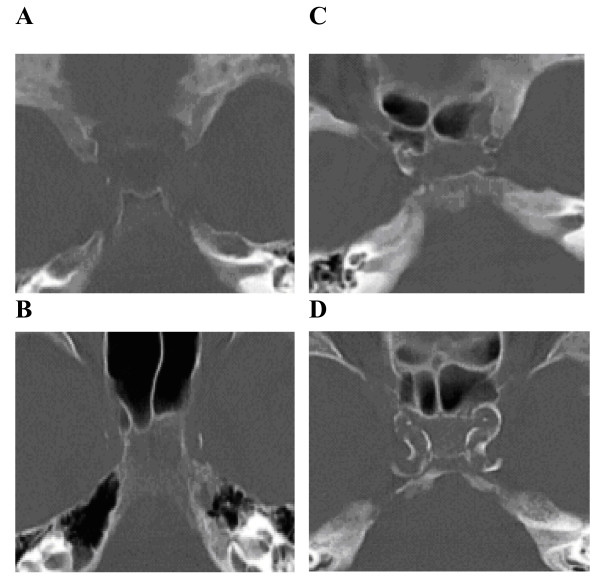
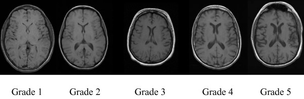
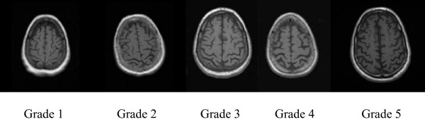
Institutional review board approval was obtained for this retrospective study incorporating 65 consecutive patients presenting acutely who had both head CT and MRI. Arterial calcifications of the intracranial cavernous carotids (ICAC) were assigned a number (1 to 4) in the bone window images from CT scans. These 4 groups were then combined into high (grades 3 and 4) and low calcium (grades 1 and 2) subgroups. Brain MRI was independently evaluated to identify cortical and central atrophy. Demographics and cardiovascular risk factors were evaluated in subjects with high and low ICAC. Relationship between CT demonstrated ICAC and brain atrophy patterns were evaluated both without and with adjustment for cerebral ischemic scores and cardiovascular risk factors.
Forty-six of the 65 (71%) patients had high ICAC on head CT. Subjects with high ICAC were older, and had higher prevalence of hypertension, diabetes, coronary artery disease (CAD), atrial fibrillation and history of previous stroke (CVA) compared to those with low ICAC. Age demonstrated strong correlation with both supratentorial atrophy patterns. There was no correlation between ICAC and cortical atrophy. There was correlation however between central atrophy and ICAC. This persisted even after adjustment for age.
Age is the most important determinant of atrophic cerebral changes. However, high ICAC demonstrated age independent association with central atrophy.
我们的目的是研究头部CT上观察到的海绵窦颈动脉钙化(ICAC)所测量的颅内动脉粥样硬化与MRI显示的幕上脑萎缩变化之间的关联。
本回顾性研究获得机构审查委员会批准,纳入65例急性就诊且同时进行了头部CT和MRI检查的连续患者。在CT扫描的骨窗图像中,颅内海绵窦颈动脉的动脉钙化(ICAC)被赋予一个数字(1至4)。然后将这4组合并为高钙(3级和4级)和低钙(1级和2级)亚组。对脑MRI进行独立评估以确定皮质和中枢萎缩。对高ICAC和低ICAC的受试者评估人口统计学和心血管危险因素。在不调整和调整脑缺血评分及心血管危险因素的情况下,评估CT显示的ICAC与脑萎缩模式之间的关系。
65例患者中有46例(71%)头部CT显示高ICAC。与低ICAC患者相比,高ICAC患者年龄更大,高血压、糖尿病、冠状动脉疾病(CAD)、心房颤动和既往中风(CVA)病史的患病率更高。年龄与幕上萎缩模式均呈强相关性。ICAC与皮质萎缩之间无相关性。然而,中枢萎缩与ICAC之间存在相关性。即使在调整年龄后,这种相关性仍然存在。
年龄是脑萎缩变化的最重要决定因素。然而,高ICAC显示出与中枢萎缩存在独立于年龄的关联。