Stilla Randall, Hanna Rebecca, Hu Xiaoping, Mariola Erica, Deshpande Gopikrishna, Sathian K
Department of Neurology, Emory University School of Medicine, Atlanta, GA 30322, USA.
J Vis. 2008 Dec 17;8(10):13.1-19. doi: 10.1167/8.10.13.
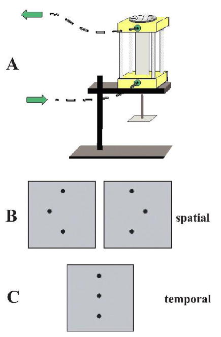
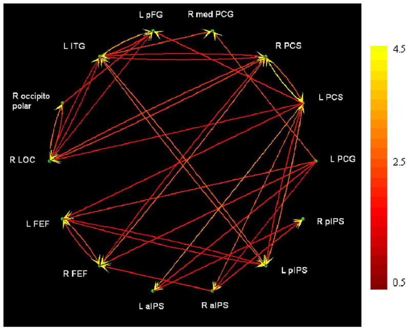
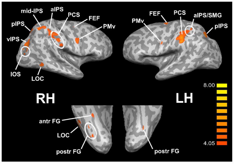
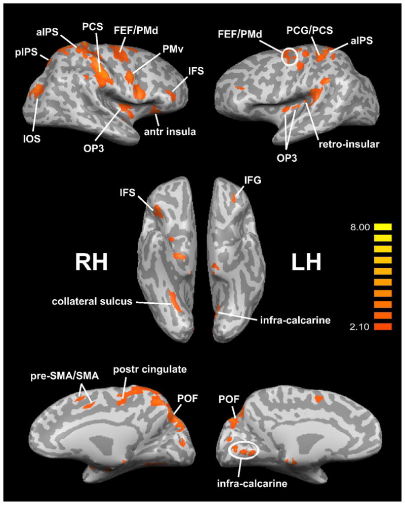
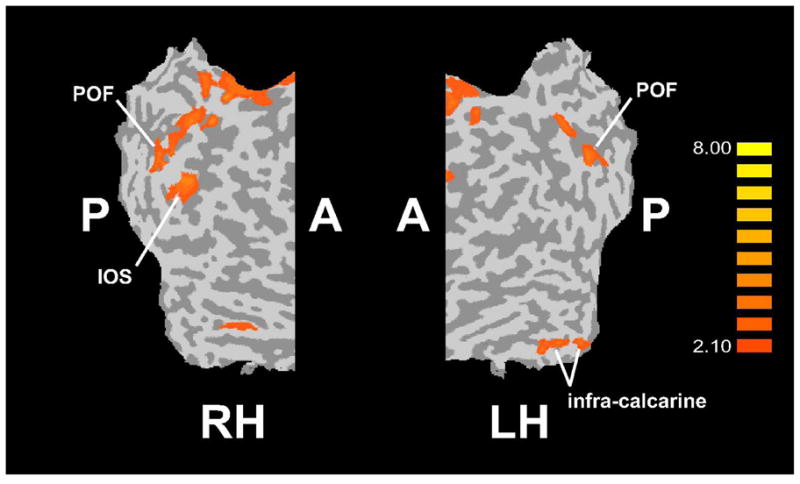
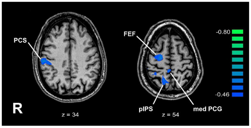
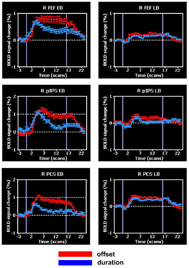
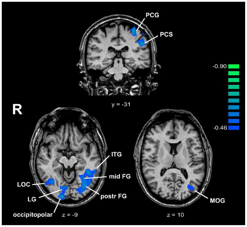
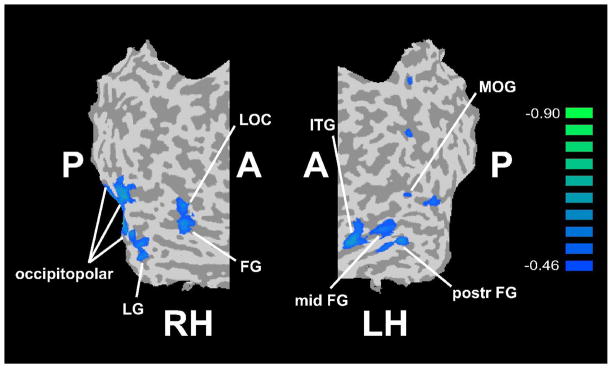
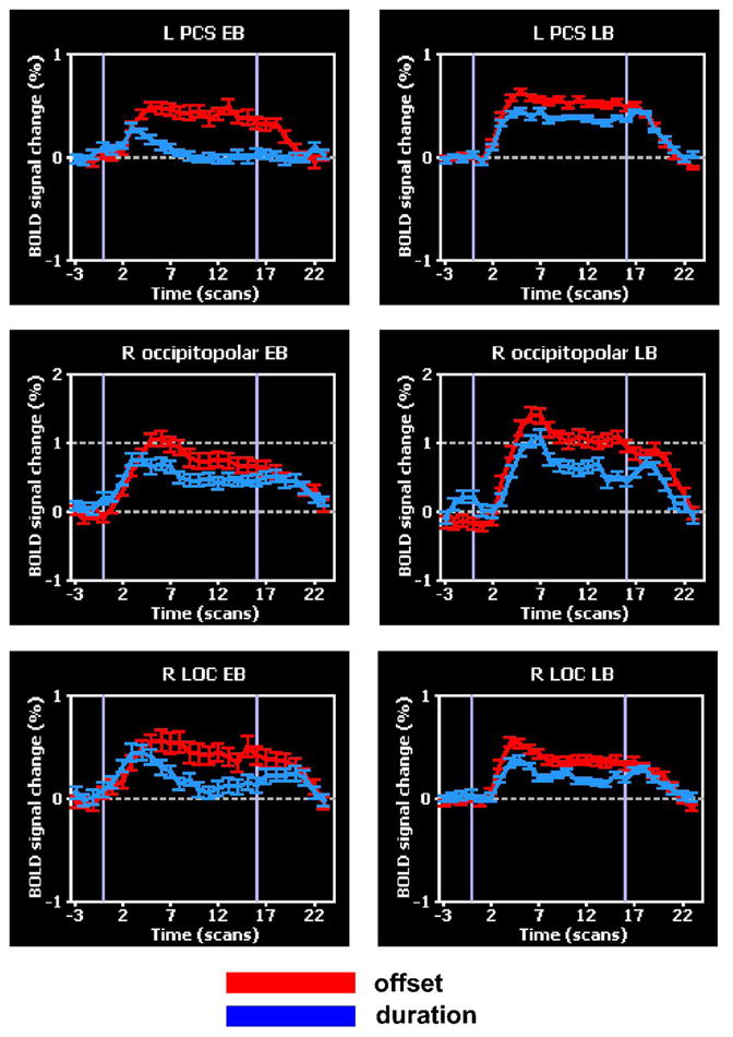
Although blindness alters neocortical processing of non-visual tasks, previous studies do not allow clear conclusions about purely perceptual tasks. We used functional magnetic resonance imaging (fMRI) to examine the neural processing underlying tactile microspatial discrimination in the blind. Activity during the tactile microspatial task was contrasted against that during a tactile temporal discrimination task. The spatially selective network included frontoparietal and visual cortical regions. Activation magnitudes in left primary somatosensory cortex and in visual cortical foci predicted acuity thresholds. Effective connectivity was investigated using multivariate Granger causality analyses. Bilateral primary somatosensory cortical foci and a left inferior temporal focus were important sources of connections. Visual cortical regions interacted mainly with one another and with somatosensory cortical regions. Among a set of distributed cortical regions exhibiting greater spatial selectivity in early blind compared to late blind individuals, the age of complete blindness was predicted by activity in a subset of frontoparietal regions and by the weight of a path from the right lateral occipital complex to right occipitopolar cortex. Thus, many aspects of neural processing during tactile microspatial discrimination differ between the blind and sighted, with some of the key differences reflecting visual cortical engagement in the blind.
虽然失明会改变新皮层对非视觉任务的处理,但先前的研究无法就纯感知任务得出明确结论。我们使用功能磁共振成像(fMRI)来研究盲人触觉微空间辨别背后的神经处理过程。将触觉微空间任务期间的活动与触觉时间辨别任务期间的活动进行对比。空间选择性网络包括额顶叶和视觉皮层区域。左侧初级体感皮层和视觉皮层病灶的激活强度可预测敏锐度阈值。使用多变量格兰杰因果分析来研究有效连接性。双侧初级体感皮层病灶和左侧颞下病灶是重要的连接源。视觉皮层区域主要相互作用,并与体感皮层区域相互作用。在一组与晚期盲人相比在早期盲人中表现出更大空间选择性的分布式皮层区域中,完全失明的年龄可通过额顶叶区域子集中的活动以及从右侧枕叶复合体到右侧枕极皮层的一条路径的权重来预测。因此,盲人在触觉微空间辨别过程中的神经处理的许多方面与有视力的人不同,其中一些关键差异反映了盲人视觉皮层的参与情况。