Magno Stefano, Terribile Daniela, Franceschini Gianluca, Fabbri Cristina, Chiesa Federica, Di Leone Alba, Costantini Melania, Belli Paolo, Masetti Riccardo
Department of Surgery, Breast Unit, Catholic University, Policlinico "A, Gemelli", Largo Agostino Gemelli, Rome, Italy.
J Med Case Rep. 2009 Jan 30;3:43. doi: 10.1186/1752-1947-3-43.
Lactating adenoma is a benign condition, representing the most prevalent breast lesion in pregnant women and during puerperium; in this paper, a case of a woman with lactating adenoma occurring during the first trimester of pregnancy is reported. There have been no reports in the literature, according to our search, focusing on magnetic resonance imaging findings in cases of lactating adenomas. Also the early onset of the lesion during the first trimester of pregnancy is quite unusual and possibly unique.
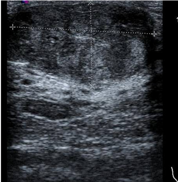
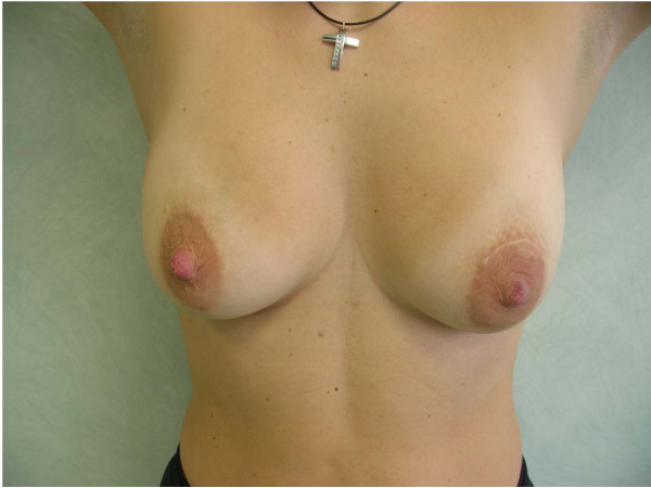
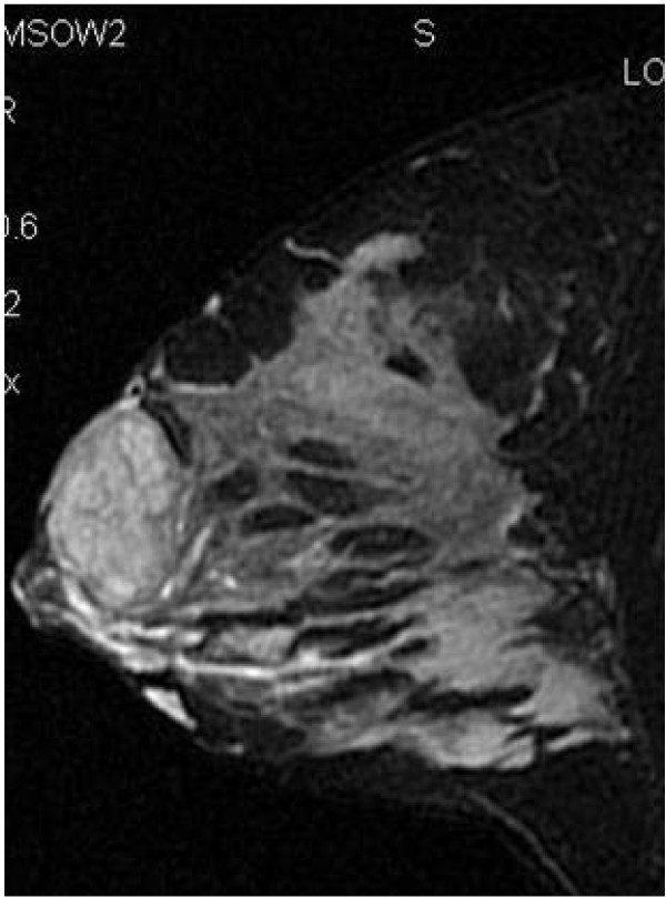
We report the case of a primiparous 30-year-old Caucasian woman, who noted an asymptomatic lump within her left breast during the 9th week of gestation, slightly increasing in size over the next few weeks. Ultrasound demonstrated a hypoecoic solid mass, hypervascularized and measuring 4 cm. On magnetic resonance imaging, performed in the first month after delivery, the lesion appeared as an ovoidal homogeneous mass, with regular margins and a significant contrast enhancement indicative of a giant adenoma.
Magnetic resonance imaging could play an important role in the differential diagnosis of pregnancy-related breast lumps, particularly during puerperium, thus avoiding unnecessary surgical biopsies.
哺乳期腺瘤是一种良性疾病,是孕妇和产褥期最常见的乳腺病变;本文报道了一例在妊娠早期发生哺乳期腺瘤的女性病例。据我们检索,文献中尚无关于哺乳期腺瘤磁共振成像表现的报道。而且该病变在妊娠早期发病相当罕见,可能是独一无二的。
我们报告一例30岁初产白种女性病例,她在妊娠第9周时发现左乳有一个无症状肿块,在接下来的几周内大小略有增加。超声显示为低回声实性肿块,血流丰富,大小为4厘米。产后第一个月进行的磁共振成像显示,病变表现为椭圆形均匀肿块,边缘规则,有明显的对比增强,提示为巨大腺瘤。
磁共振成像在妊娠相关乳腺肿块的鉴别诊断中可发挥重要作用,尤其是在产褥期,从而避免不必要的手术活检。