Nguyen David, Deng Ping, Matthews Elizabeth A, Kim Doo-Sik, Feng Guoping, Dickenson Anthony H, Xu Zao C, Luo Z David
Department of Anesthesiology & Perioperative Care, School of Medicine, University of California Irvine, Irvine, CA 92697, USA.
Mol Pain. 2009 Feb 12;5:6. doi: 10.1186/1744-8069-5-6.
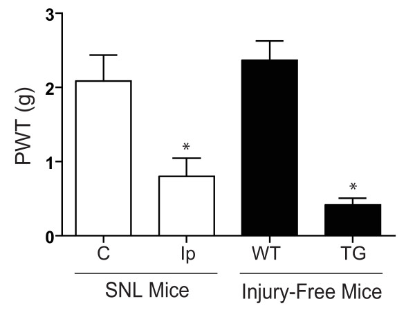
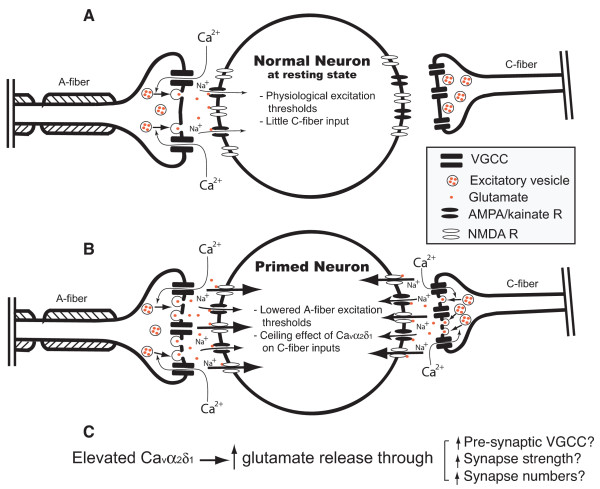
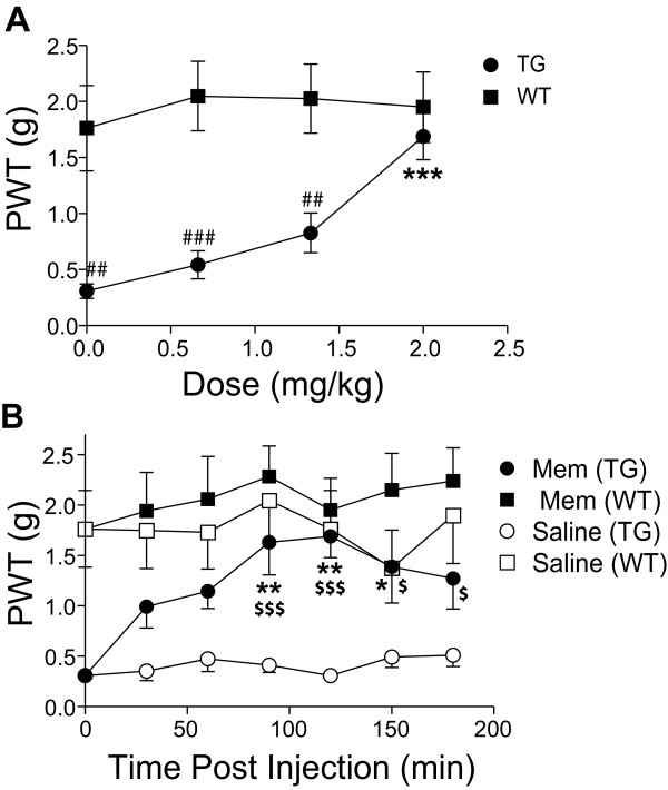
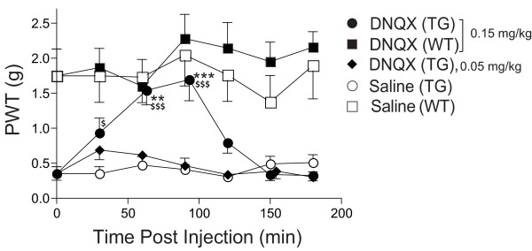
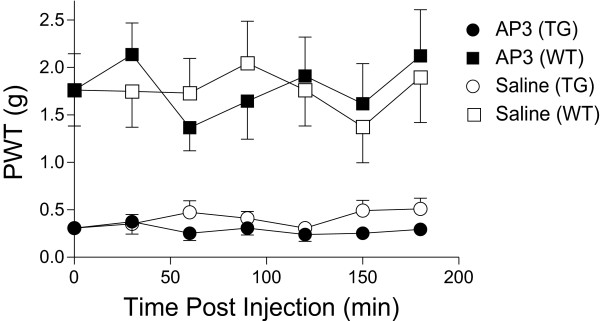
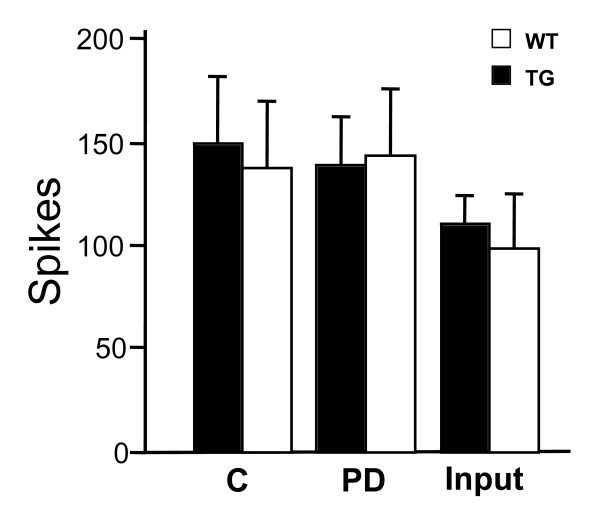
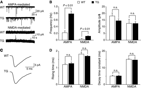
Nerve injury-induced expression of the spinal calcium channel alpha-2-delta-1 subunit (Cavalpha2delta1) has been shown to mediate behavioral hypersensitivity through a yet identified mechanism. We examined if this neuroplasticity modulates behavioral hypersensitivity by regulating spinal glutamatergic neurotransmission in injury-free transgenic mice overexpressing the Cavalpha2delta1 proteins in neuronal tissues. The transgenic mice exhibited hypersensitivity to mechanical stimulation (allodynia) similar to the spinal nerve ligation injury model. Intrathecally delivered antagonists for N-methyl-D-aspartate (NMDA) and alpha-amino-3-hydroxyl-5-methylisoxazole-4-propionic acid (AMPA)/kainate receptors, but not for the metabotropic glutamate receptors, caused a dose-dependent allodynia reversal in the transgenic mice without changing the behavioral sensitivity in wild-type mice. This suggests that elevated spinal Cavalpha2delta1 mediates allodynia through a pathway involving activation of selective glutamate receptors. To determine if this is mediated by enhanced spinal neuronal excitability or pre-synaptic glutamate release in deep-dorsal horn, we examined wide-dynamic-range (WDR) neuron excitability with extracellular recording and glutamate-mediated excitatory postsynaptic currents with whole-cell patch recording in deep-dorsal horn of the Cavalpha2delta1 transgenic mice. Our data indicated that overexpression of Cavalpha2delta1 in neuronal tissues led to increased frequency, but not amplitude, of miniature excitatory post synaptic currents mediated mainly by AMPA/kainate receptors at physiological membrane potentials, and also by NMDA receptors upon depolarization, without changing the excitability of WDR neurons to high intensity stimulation. Together, these findings support a mechanism of Cavalpha2delta1-mediated spinal sensitization in which elevated Cavalpha2delta1 causes increased pre-synaptic glutamate release that leads to reduced excitation thresholds of post-synaptic dorsal horn neurons to innocuous stimuli. This spinal sensitization mechanism may mediate at least partially the neuropathic pain states derived from increased pre-synaptic Cavalpha2delta1 expression.
神经损伤诱导的脊髓钙通道α-2-δ-1亚基(Cavα2δ1)表达已被证明可通过一种尚未明确的机制介导行为超敏反应。我们在神经元组织中过表达Cavα2δ1蛋白的无损伤转基因小鼠中研究了这种神经可塑性是否通过调节脊髓谷氨酸能神经传递来调节行为超敏反应。转基因小鼠表现出对机械刺激的超敏反应(痛觉过敏),类似于脊髓神经结扎损伤模型。鞘内注射N-甲基-D-天冬氨酸(NMDA)和α-氨基-3-羟基-5-甲基异恶唑-4-丙酸(AMPA)/海人藻酸受体拮抗剂,但不注射代谢型谷氨酸受体拮抗剂,可使转基因小鼠的痛觉过敏呈剂量依赖性逆转,而不改变野生型小鼠的行为敏感性。这表明脊髓中升高的Cavα2δ1通过涉及选择性谷氨酸受体激活的途径介导痛觉过敏。为了确定这是否由脊髓神经元兴奋性增强或背角深层突触前谷氨酸释放介导,我们在Cavα2δ1转基因小鼠的背角深层用细胞外记录检查了广动力范围(WDR)神经元兴奋性,并用全细胞膜片钳记录检查了谷氨酸介导的兴奋性突触后电流。我们的数据表明,神经元组织中Cavα2δ1的过表达导致主要由AMPA/海人藻酸受体在生理膜电位下介导的微小兴奋性突触后电流频率增加,但幅度不变,在去极化时由NMDA受体介导的微小兴奋性突触后电流频率也增加,而不改变WDR神经元对高强度刺激的兴奋性。总之,这些发现支持了Cavα2δ1介导的脊髓致敏机制,其中升高的Cavα2δ1导致突触前谷氨酸释放增加,从而导致突触后背角神经元对无害刺激的兴奋阈值降低。这种脊髓致敏机制可能至少部分介导了由突触前Cavα2δ1表达增加引起的神经性疼痛状态。