Cokakli Murat, Erdal Esra, Nart Deniz, Yilmaz Funda, Sagol Ozgul, Kilic Murat, Karademir Sedat, Atabey Nese
Dokuz Eylul University, Faculty of Medicine, Department of Medical Biology and Genetics, Inciralti, Izmir, Turkey.
BMC Cancer. 2009 Feb 24;9:65. doi: 10.1186/1471-2407-9-65.
Caveolin-1 is the main component of caveolae membrane structures and has different roles during tumorigenesis in different cancer types with varying expression profiles, indicating that the role of caveolin-1 varies according to tumor type. In this study, we investigated the role and expression of caveolin-1 in hepatocellular carcinogenesis.
We analyzed the expression of Caveolin-1 in 96 hepatocellular carcinoma (HCC), 29 cirrhosis, 20 normal liver tissues and 9 HCC cell lines by immunostaining and western blotting, respectively. After caveolin-1 was stably transfected to HepG2 and Huh7 cells, the effects of Caveolin-1 on the cellular motility, matrix invasion and anchorage-independent growth were studied. Also, caveolae structure was disrupted in endogenously caveolin expressing cells, SNU 449 and SNU 475 by addition of methyl-beta-cyclodextrin and analyzed cellular motility and invasion.
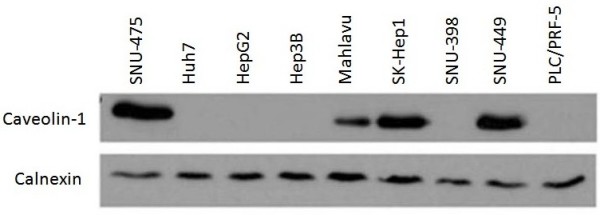
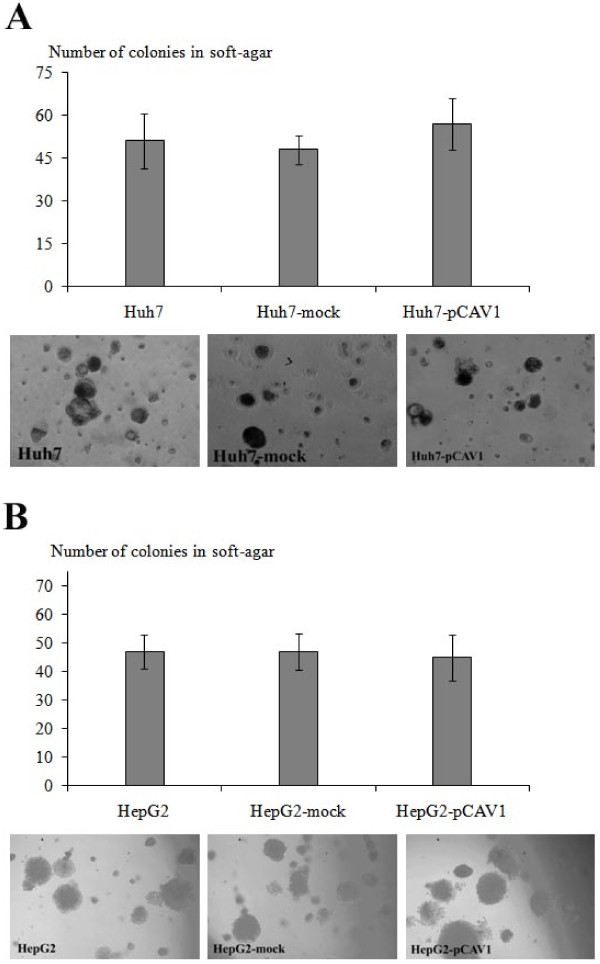
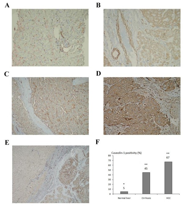
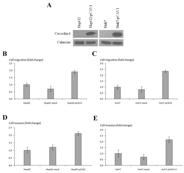
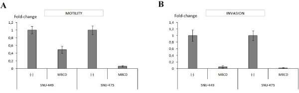
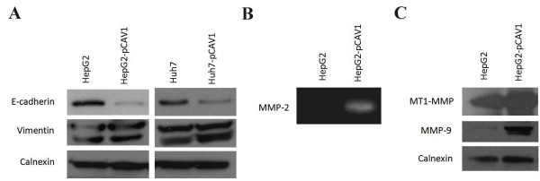
In HCC cell lines, Caveolin-1 expression is correlated to differentiation and basal motility status of these cells. The percentage of Caveolin-1 positivity was found extremely low in normal liver tissue (5%) while it was increased in cirrhosis (45%) and in HCC (66%) (p = 0.002 and p = 0.001 respectively). Cav-1 expression in poorly differentiated HCC samples has been found significantly higher than well differentiated ones (p = 0.001). The caveolin-1 expression was found significantly higher in tumor cells than its peritumoral cirrhotic tissues in HCC samples (p < 0.001). Additionally, the patients with positive staining for Caveolin-1 had significantly higher portal vein invasion than those with negative staining (p = 0.02). Caveolin-1 overexpression increased motility and invasion of HepG2 and Huh7 cells. And disruption of caveolae results in a dramatic decline in both motility and invasion abilities in SNU-449 and SNU-475 cells. Furthermore, caveolin-1 overexpression resulted in down-regulation of E-cadherin while up-regulation of Vimentin. Also, it increased secreted MMP-2 and expression levels of MMP-9 and MT1-MMP. There was no significant difference in colony formation in soft agar between stable clones and parental ones.
In conclusion, stepwise increase in Cav-1 expression in neoplastic stage with respect to pre-neoplastic stage during hepatocellular carcinogenesis and its ability to stimulate HCC cell motility and invasiveness indicate that this protein plays a crucial role in tumor progression.
小窝蛋白-1是小窝膜结构的主要成分,在不同癌症类型的肿瘤发生过程中具有不同作用,其表达谱各异,这表明小窝蛋白-1的作用因肿瘤类型而异。在本研究中,我们调查了小窝蛋白-1在肝细胞癌发生中的作用和表达情况。
我们分别通过免疫染色和蛋白质印迹法分析了96例肝细胞癌(HCC)、29例肝硬化、20例正常肝组织和9种HCC细胞系中小窝蛋白-1的表达。将小窝蛋白-1稳定转染至HepG2和Huh7细胞后,研究了小窝蛋白-1对细胞运动性、基质侵袭和非锚定依赖性生长的影响。此外,通过添加甲基-β-环糊精破坏内源性表达小窝蛋白的细胞SNU 449和SNU 475中的小窝结构,并分析细胞运动性和侵袭能力。
在HCC细胞系中,小窝蛋白-1的表达与这些细胞的分化和基础运动状态相关。在正常肝组织中,小窝蛋白-1阳性率极低(5%),而在肝硬化组织中升高(45%),在HCC组织中更高(66%)(分别为p = 0.002和p = 0.001)。在低分化HCC样本中,小窝蛋白-1的表达明显高于高分化样本(p = 0.001)。在HCC样本中,肿瘤细胞中的小窝蛋白-1表达明显高于其瘤旁肝硬化组织(p < 0.001)。此外,小窝蛋白-1染色阳性的患者门静脉侵犯明显高于染色阴性的患者(p = 0.02)。小窝蛋白-1过表达增加了HepG2和Huh7细胞的运动性和侵袭能力。小窝结构的破坏导致SNU-449和SNU-475细胞运动性和侵袭能力显著下降。此外,小窝蛋白-1过表达导致E-钙黏蛋白下调,波形蛋白上调。同时,它增加了MMP-2的分泌以及MMP-9和MT1-MMP的表达水平。稳定克隆与亲代细胞在软琼脂中的集落形成没有显著差异。
总之,在肝细胞癌发生过程中,与癌前阶段相比,肿瘤阶段小窝蛋白-1表达逐步增加,且其具有刺激HCC细胞运动性和侵袭性的能力,这表明该蛋白在肿瘤进展中起关键作用。