Department of Molecular and System Pharmacology, Graduate School of Pharmaceutical Sciences, Kyushu University, Fukuoka, Japan.
J Cell Mol Med. 2009 Sep;13(9B):3251-9. doi: 10.1111/j.1582-4934.2009.00719.x. Epub 2009 Feb 27.
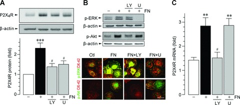
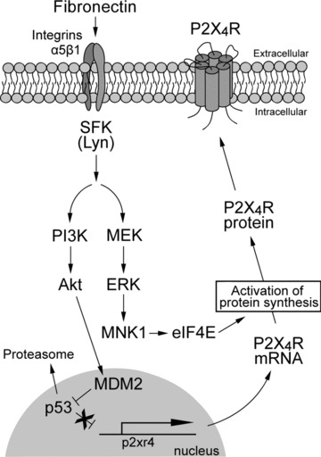
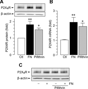
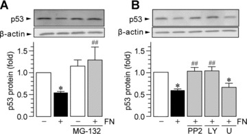
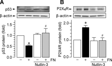
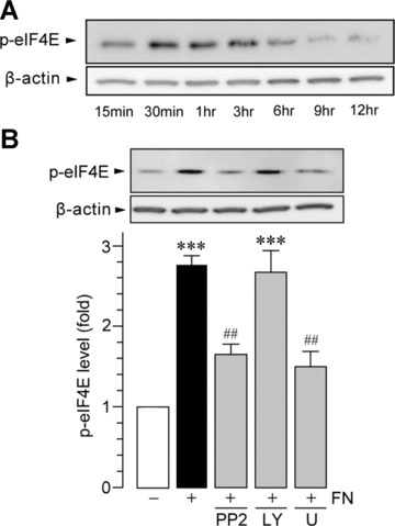
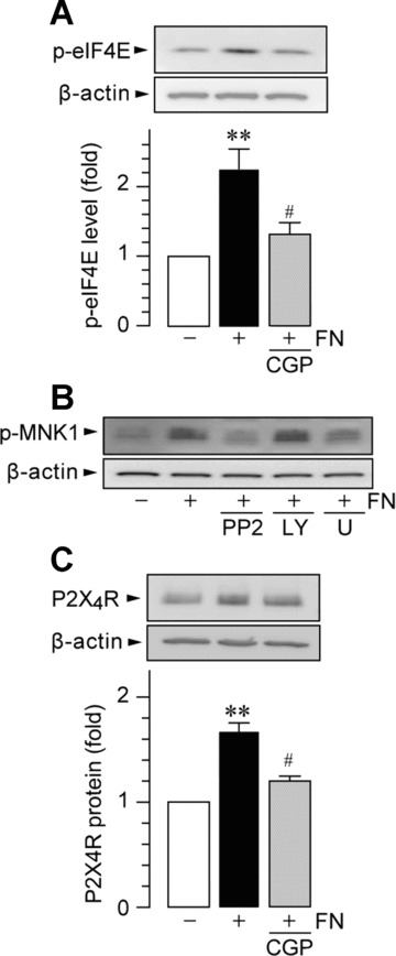
Microglia are resident immune cells in the central nervous system that become activated and produce pro-inflammatory and neurotrophic factors upon activation of various cell-surface receptors. The P2X(4) receptor (P2X(4)R) is a sub-type of the purinergic ion-channel receptors expressed in microglia. P2X(4)R expression is up-regulated under inflammatory or neurodegenerative conditions, and this up-regulation is implicated in disease pathology. However, the molecular mechanism underlying up-regulation of P2X(4)R in microglia remains unknown. In the present study, we investigated the intracellular signal transduction pathway that promotes P2X(4)R expression in microglia in response to fibronectin, an extracellular matrix protein that has previously been shown to stimulate P2X(4)R expression. We found that in fibronectin-stimulated microglia, activation of phosphatidylinositol 3-kinase (PI3K)-Akt and mitogen-activated protein kinase kinase (MAPK kinase, MEK)-extracellular signal-regulated kinase (ERK) signalling cascades occurred divergently downstream of Src-family kinases (SFKs). Pharmacological interference of PI3K-Akt signalling inhibited fibronectin-induced P2X(4)R gene expression. Activation of PI3K-Akt signalling resulted in a decrease in the protein level of the transcription factor p53 via mouse double minute 2 (MDM2), an effect that was prevented by MG-132, an inhibitor of the proteasome. In microglia pre-treated with MG-132, fibronectin failed to up-regulate P2X(4)R expression. Conversely, an inhibitor of p53 caused increased expression of P2X(4)R, implying a negative regulatory role of p53. On the other hand, inhibiting MEK-ERK signalling activated by fibronectin suppressed an increase in P2X(4)R protein but interestingly did not affect the level of P2X(4)R mRNA. We also found that fibronectin stimulation resulted in the activation of the translational factor eIF4E via MAPK-interacting protein kinase-1 (MNK1) in an MEK-ERK signalling-dependent manner, and an MNK1 inhibitor attenuated the increase in P2X(4)R protein. Together, these results suggest that the PI3K-Akt and MEK-ERK signalling cascades have distinct roles in the up-regulation of P2X(4)R expression in microglia at transcriptional and post-transcriptional levels, respectively.
小胶质细胞是中枢神经系统中的固有免疫细胞,在各种细胞表面受体被激活后,会产生促炎和神经营养因子。P2X(4)受体(P2X(4)R)是嘌呤能离子通道受体的一种亚型,在小胶质细胞中表达。在炎症或神经退行性条件下,P2X(4)R 的表达上调,这种上调与疾病病理有关。然而,小胶质细胞中 P2X(4)R 上调的分子机制尚不清楚。在本研究中,我们研究了促进纤维连接蛋白刺激的小胶质细胞中 P2X(4)R 表达的细胞内信号转导途径,纤维连接蛋白是一种先前已被证明可刺激 P2X(4)R 表达的细胞外基质蛋白。我们发现,在纤维连接蛋白刺激的小胶质细胞中,Src 家族激酶(SFKs)下游的磷酸肌醇 3-激酶(PI3K)-Akt 和丝裂原活化蛋白激酶激酶(MAPK 激酶,MEK)-细胞外信号调节激酶(ERK)信号级联反应以不同的方式发生。PI3K-Akt 信号通路的药理学干扰抑制了纤维连接蛋白诱导的 P2X(4)R 基因表达。PI3K-Akt 信号通路的激活导致转录因子 p53 的蛋白水平通过鼠双微体 2(MDM2)降低,这种作用被蛋白酶体抑制剂 MG-132 所阻止。在用 MG-132 预处理的小胶质细胞中,纤维连接蛋白未能上调 P2X(4)R 的表达。相反,p53 的抑制剂导致 P2X(4)R 的表达增加,表明 p53 具有负调节作用。另一方面,抑制纤维连接蛋白激活的 MEK-ERK 信号通路抑制了 P2X(4)R 蛋白的增加,但有趣的是,它不影响 P2X(4)R mRNA 的水平。我们还发现,纤维连接蛋白刺激通过 MAPK 相互作用蛋白激酶-1(MNK1)以 MEK-ERK 信号依赖性方式导致翻译因子 eIF4E 的激活,MNK1 抑制剂减弱了 P2X(4)R 蛋白的增加。总之,这些结果表明,PI3K-Akt 和 MEK-ERK 信号级联在转录和转录后水平分别对小胶质细胞中 P2X(4)R 表达的上调具有不同的作用。