French Amanda D, Fiori Jennifer L, Camilli Tura C, Leotlela Poloko D, O'Connell Michael P, Frank Brittany P, Subaran Sarah, Indig Fred E, Taub Dennis D, Weeraratna Ashani T
Laboratory of Immunology, National Institute on Aging, Baltimore, MD 21124, USA.
Int J Med Sci. 2009;6(2):93-101. doi: 10.7150/ijms.6.93. Epub 2009 Mar 12.
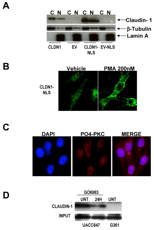
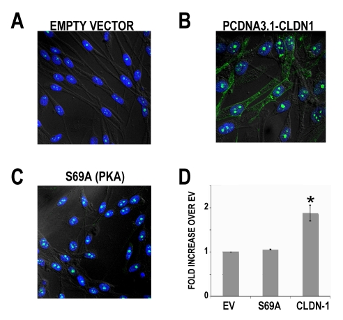
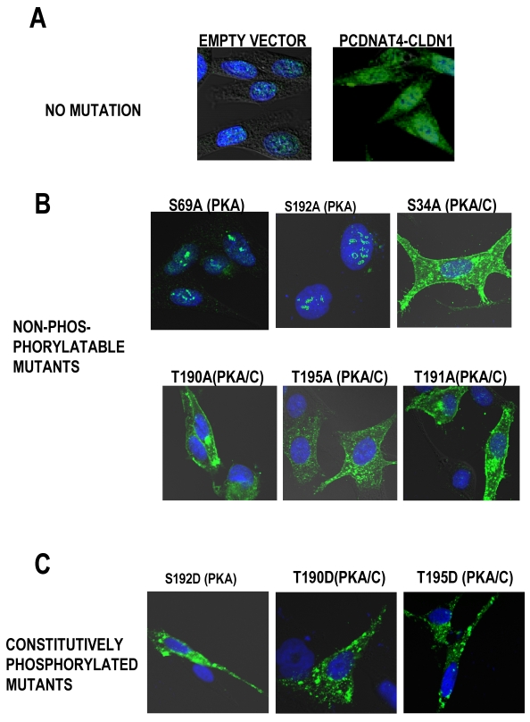
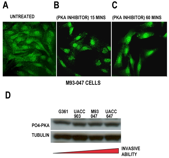
Cytoplasmic expression of claudin-1 in metastatic melanoma cells correlates to increased migration, and increased secretion of MMP-2 in a PKC dependent manner, whereas claudin-1 nuclear expression is found in benign nevi. Melanoma cells were transfected with a vector expressing CLDN-1 fused to a nuclear localization signal (NLS). Despite significant nuclear localization of claudin-1, there was still transport of claudin-1 to the cytoplasm. Phorbol ester treatment of cells transfected with NLS-claudin-1 resulted in an exclusion of claudin-1 from the nucleus, despite the NLS. To ascertain whether PKC or PKA were involved in this translocation, we mutated the putative phosphorylation sites within the protein. We found that mutating the PKC phosphorylation sites to mimic a non-phosphorylated state did not cause a shift of claudin-1 to the nucleus of the cells, but mutating the PKA sites did. Mutations of either site to mimic constitutive phosphorylation resulted in cytoplasmic claudin-1 expression. Stable claudin-1 transfectants containing non-phosphorylatable PKA sites exhibited decreased motility. These data imply that subcellular localization of claudin-1 can be controlled by phosphorylation, dicating effects on metastatic capacity.
紧密连接蛋白-1(claudin-1)在转移性黑色素瘤细胞中的细胞质表达与迁移增加以及基质金属蛋白酶-2(MMP-2)以蛋白激酶C(PKC)依赖方式分泌增加相关,而紧密连接蛋白-1的核表达则见于良性痣。黑色素瘤细胞用表达与核定位信号(NLS)融合的CLDN-1的载体进行转染。尽管紧密连接蛋白-1有明显的核定位,但仍有紧密连接蛋白-1转运至细胞质。用佛波酯处理转染了NLS-紧密连接蛋白-1的细胞,尽管有NLS,紧密连接蛋白-1仍被排除在细胞核外。为确定PKC或蛋白激酶A(PKA)是否参与这种易位,我们对该蛋白内的假定磷酸化位点进行了突变。我们发现,将PKC磷酸化位点突变为模拟非磷酸化状态不会导致紧密连接蛋白-1向细胞核移位,但将PKA位点突变则会。将任一位点突变为模拟组成型磷酸化会导致紧密连接蛋白-1在细胞质中表达。含有不可磷酸化PKA位点的稳定紧密连接蛋白-1转染子的运动性降低。这些数据表明紧密连接蛋白-1的亚细胞定位可通过磷酸化来控制,这表明其对转移能力有影响。