Nishikimi Akihiko, Fukuhara Hideo, Su Wenjuan, Hongu Tsunaki, Takasuga Shunsuke, Mihara Hisashi, Cao Qinhong, Sanematsu Fumiyuki, Kanai Motomu, Hasegawa Hiroshi, Tanaka Yoshihiko, Shibasaki Masakatsu, Kanaho Yasunori, Sasaki Takehiko, Frohman Michael A, Fukui Yoshinori
Division of Immunogenetics, Department of Immunobiology and Neuroscience, Medical Institute of Bioregulation, Kyushu University, Fukuoka 812-8582, Japan.
Science. 2009 Apr 17;324(5925):384-7. doi: 10.1126/science.1170179. Epub 2009 Mar 26.
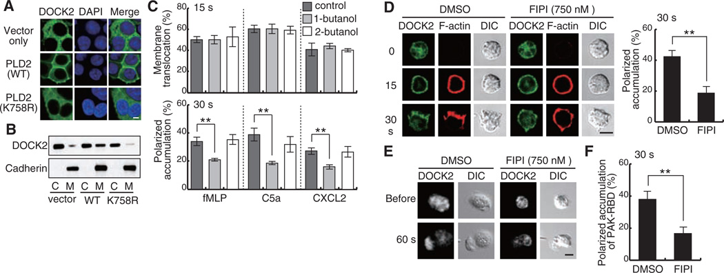
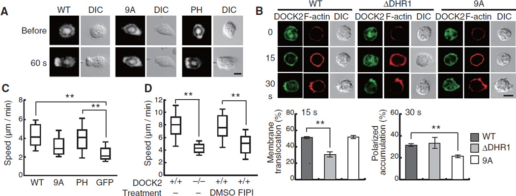
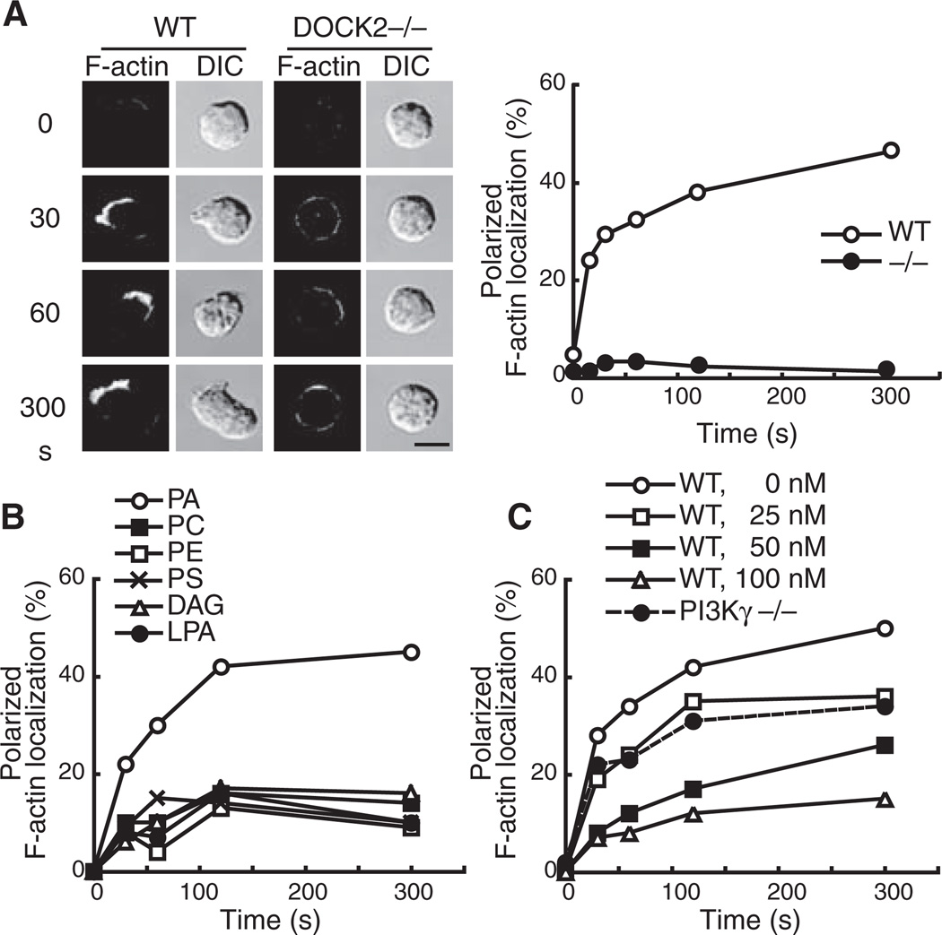
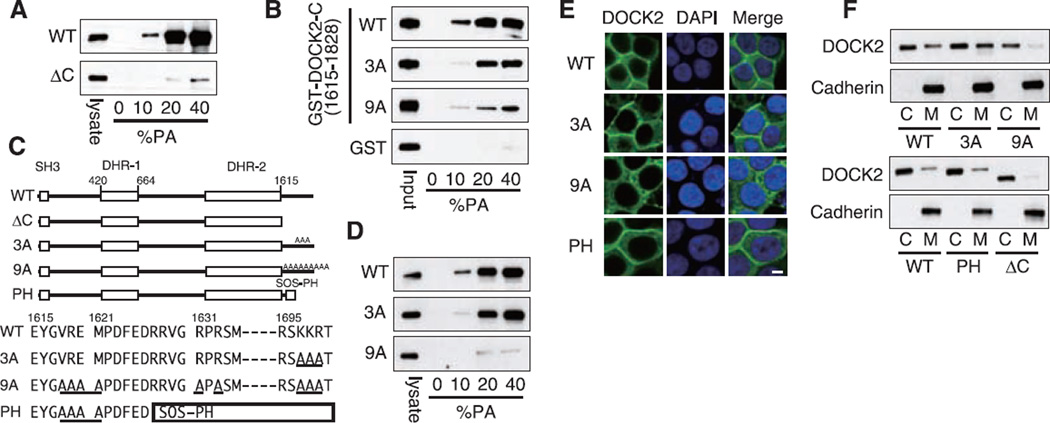
During chemotaxis, activation of the small guanosine triphosphatase Rac is spatially regulated to organize the extension of membrane protrusions in the direction of migration. In neutrophils, Rac activation is primarily mediated by DOCK2, an atypical guanine nucleotide exchange factor. Upon stimulation, we found that DOCK2 rapidly translocated to the plasma membrane in a phosphatidylinositol 3,4,5-trisphosphate-dependent manner. However, subsequent accumulation of DOCK2 at the leading edge required phospholipase D-mediated synthesis of phosphatidic acid, which stabilized DOCK2 there by means of interaction with a polybasic amino acid cluster, resulting in increased local actin polymerization. When this interaction was blocked, neutrophils failed to form leading edges properly and exhibited defects in chemotaxis. Thus, intracellular DOCK2 dynamics are sequentially regulated by distinct phospholipids to localize Rac activation during neutrophil chemotaxis.
在趋化作用过程中,小GTP酶Rac的激活在空间上受到调控,以在迁移方向上组织膜突起的延伸。在中性粒细胞中,Rac激活主要由非典型鸟嘌呤核苷酸交换因子DOCK2介导。受到刺激后,我们发现DOCK2以磷脂酰肌醇3,4,5 -三磷酸依赖性方式迅速转运至质膜。然而,DOCK2随后在前缘的积累需要磷脂酶D介导的磷脂酸合成,磷脂酸通过与多碱性氨基酸簇相互作用将DOCK2稳定在那里,导致局部肌动蛋白聚合增加。当这种相互作用被阻断时,中性粒细胞无法正常形成前缘,并在趋化作用中表现出缺陷。因此,在中性粒细胞趋化作用过程中,细胞内DOCK2的动态变化由不同的磷脂顺序调节,以定位Rac激活。