Department of Surgery, University of Washington, Seattle, Washington, USA.
Shock. 2009 Dec;32(6):572-7. doi: 10.1097/SHK.0b013e3181a72530.
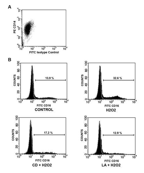
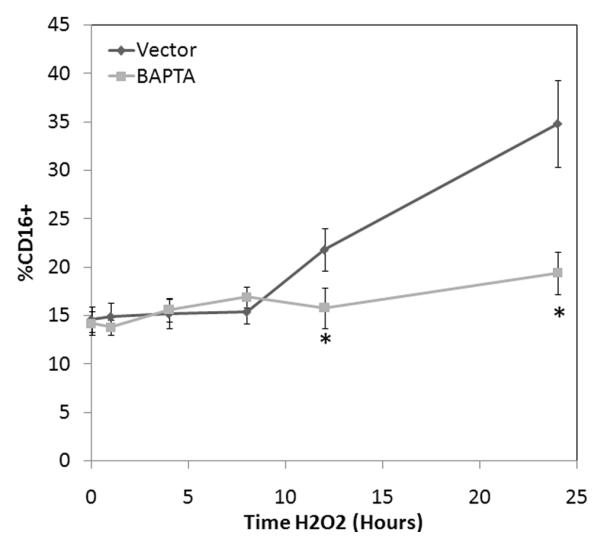
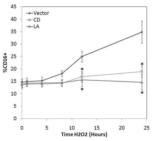
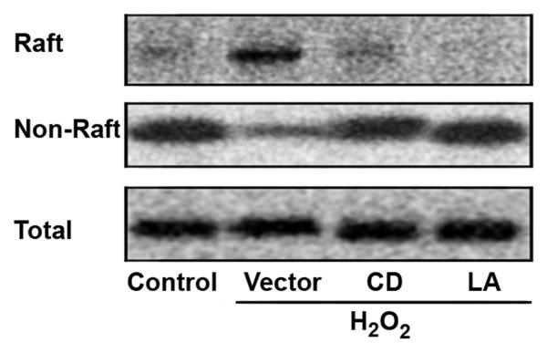
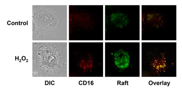
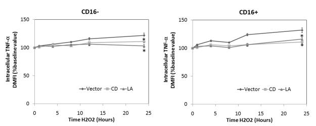
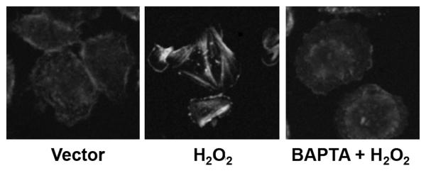
Oxidative stress during reperfusion of ischemia is associated with a phenotypic change in circulating monocytes from CD14++CD16- to a proinflammatory CD14+CD16+ subpopulation resulting in altered immunity and development of organ failure. However, the mechanism responsible remains unknown. We hypothesize that this phenotypic change, modeled by hydrogen peroxide exposure in vitro, is due to oxidative-induced intracellular calcium flux and distinct cytoskeletal and lipid raft changes. Peripheral blood monocytes obtained from healthy volunteers underwent 100 mM H2O2 exposure for 0 to 24 h. Selected cells were pretreated with 2 microM cytochalasin D, 1 microM lactrunculin A, or 30 microM 1,2-bis(2-aminophenoxy)ethane-N,N,N',N'-tetraacetic acid for 30 min. Cells underwent fluorescence-activated cell sorter for CD14, CD16, and cytokine expression. Cellular and lipid raft CD16 expression was determined by immunoblot and confocal microscopy. H2O2 exposed monocytes underwent a rapid time-dependent increase in the surface expression of CD16 from 12.81% +/- 3.53% to 37.12% +/- 7.61% at 24 h (P = 0.001). Total cellular CD16 was not changed by H2O2, but an increase in lipid raft and decrease in intracellular CD16 expression were seen after H2O2 exposure. This increase in CD16 expression was associated with a 27% increase in intracellular TNF-alpha, an alteration in actin polymerization, and the formation of raft macrodomains. These changes induced by H2O2 were inhibited by inhibition of actin polymerization (cytochalasin D and lactrunculin A) and intracellular calcium flux [1,2-bis(2-aminophenoxy)ethane-N,N,N',N'-tetraacetic acid]. This study provides the first evidence that phenotypic alterations induced by oxidative stress during reperfusion may occur as a result of changes in cytoskeletal architecture due to calcium flux that result in lipid raft alterations rather than solely from demargination and/or production of bone marrow-derived CD16+ monocytes.
再灌注期间的氧化应激与循环单核细胞从 CD14++CD16-向促炎的 CD14+CD16+亚群的表型变化有关,导致免疫改变和器官衰竭的发生。然而,其具体机制尚不清楚。我们假设这种表型变化,通过体外过氧化氢暴露来模拟,是由于氧化诱导的细胞内钙流以及不同的细胞骨架和脂质筏变化所致。从健康志愿者中获得外周血单核细胞,用 100mM H2O2 孵育 0 至 24 小时。选择的细胞用 2μM 细胞松弛素 D、1μM 拉曲库林 A 或 30μM 1,2-双(2-氨基苯氧基)乙烷-N,N,N',N'-四乙酸预处理 30 分钟。细胞经荧光激活细胞分选仪进行 CD14、CD16 和细胞因子表达检测。细胞和脂质筏 CD16 的表达通过免疫印迹和共聚焦显微镜确定。H2O2 暴露的单核细胞表面 CD16 的表达迅速随时间增加,在 24 小时时从 12.81%±3.53%增加至 37.12%±7.61%(P=0.001)。H2O2 未改变总细胞 CD16,但 H2O2 暴露后可见脂质筏 CD16 增加和细胞内 CD16 表达减少。这种 CD16 表达的增加与细胞内 TNF-α增加 27%、肌动蛋白聚合改变和筏状大分子域的形成有关。H2O2 诱导的这些变化可通过抑制肌动蛋白聚合(细胞松弛素 D 和拉曲库林 A)和细胞内钙流[1,2-双(2-氨基苯氧基)乙烷-N,N,N',N'-四乙酸]而被抑制。本研究首次提供证据表明,再灌注期间氧化应激诱导的表型改变可能是由于钙流引起的细胞骨架结构改变导致脂质筏改变所致,而不仅仅是由于边缘去除和/或骨髓衍生 CD16+单核细胞的产生。