Ernst Thomas, Yakupov Renat, Nakama Helenna, Crocket Grace, Cole Michael, Watters Michael, Ricardo-Dukelow Mary Lynn, Chang Linda
Department of Medicine, University of Hawaii at Manoa, and the Queen's Medical Center, Honolulu, HI 96813, USA.
Ann Neurol. 2009 Mar;65(3):316-25. doi: 10.1002/ana.21594.
To determine whether brain activation changes in clinically and neurocognitively normal human immunodeficiency virus (HIV)-infected and in HIV-seronegative control (SN) participants over a 1-year period.
Functional magnetic resonance imaging (fMRI) was performed in 32 SN and 31 HIV patients (all with stable combination antiretroviral treatment) at baseline and after 1 year. Each participant performed a set of visual attention tasks with increasing attentional load (from tracking two, three, or four balls). All HIV and SN participants had normal neuropsychological function at both examinations.
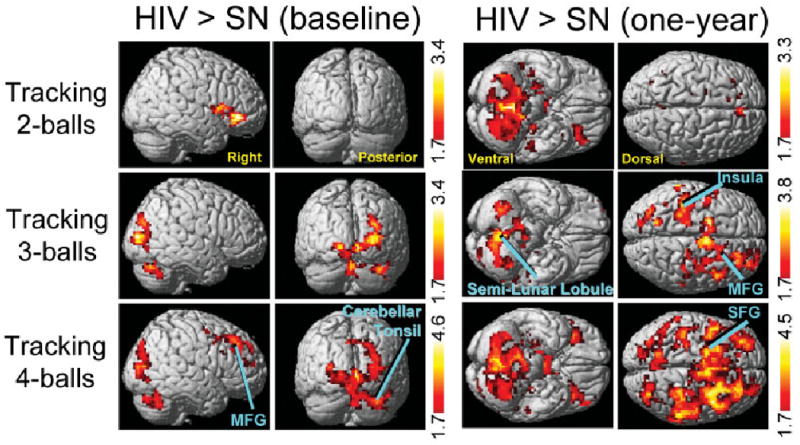
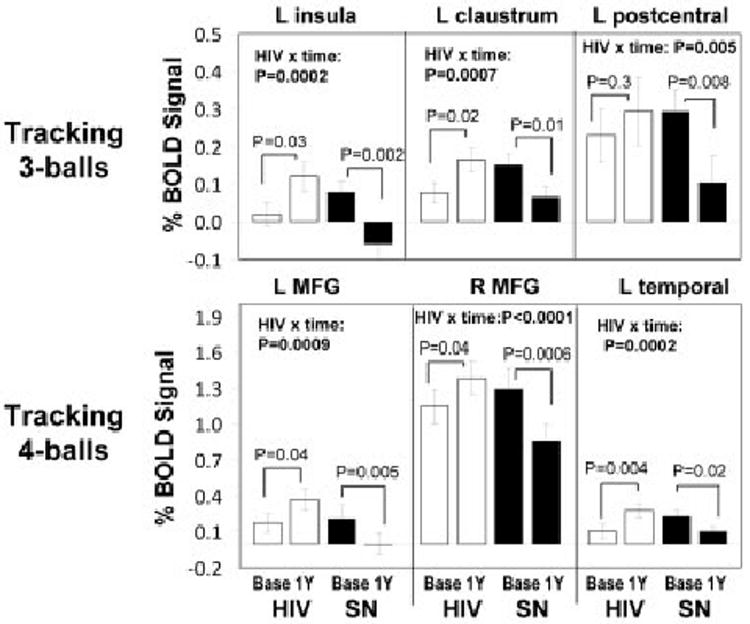
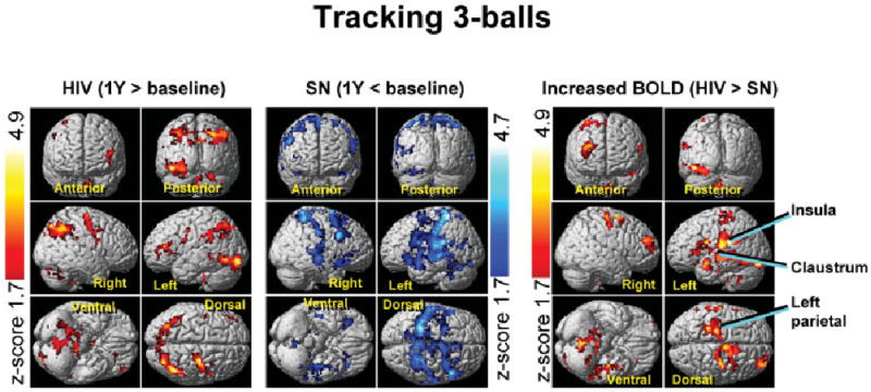
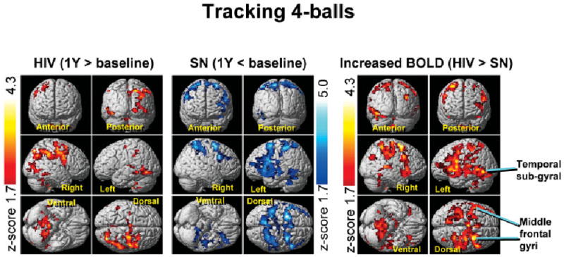
Over 1 year, HIV patients showed no change in their neurocognitive status or in task performance during fMRI. However, HIV patients showed significant 1-year increases in fMRI signals in the prefrontal and posterior parietal cortices for the more difficult tasks, whereas SN control participants showed only decreases in brain activation in these regions. This resulted in significant interactions between HIV status and time of study in left insula, left parietal, left temporal, and several frontal regions (left and right middle frontal gyrus, and anterior cingulate).
Because fMRI task performance remained unchanged in both groups, the HIV patients appeared to maintain performance by increasing usage of the attention network, whereas the control participants reduced usage of the attention network after 1 year. These findings suggest improved efficiency or a practice effect in the SN participants but declined efficiency of the neural substrate in HIV patients, possibly because of ongoing brain injury associated with the HIV infection, despite their apparent stable clinical course.
确定在1年时间里,临床和神经认知功能正常的人类免疫缺陷病毒(HIV)感染者及HIV血清学阴性对照(SN)参与者的大脑激活变化情况。
对32名SN参与者和31名HIV患者(均接受稳定的联合抗逆转录病毒治疗)在基线时和1年后进行功能磁共振成像(fMRI)检查。每位参与者执行一组视觉注意力任务,注意力负荷逐渐增加(从跟踪两个、三个或四个球开始)。所有HIV和SN参与者在两次检查时神经心理功能均正常。
在1年时间里,HIV患者在fMRI期间神经认知状态和任务表现均无变化。然而,对于更困难的任务,HIV患者在额叶和顶叶后部皮质的fMRI信号在1年中有显著增加,而SN对照参与者在这些区域的大脑激活仅出现减少。这导致在左侧岛叶、左侧顶叶、左侧颞叶以及几个额叶区域(左侧和右侧额中回以及前扣带回),HIV状态与研究时间之间存在显著交互作用。
由于两组的fMRI任务表现均未改变,HIV患者似乎通过增加注意力网络的使用来维持表现,而对照参与者在1年后减少了注意力网络的使用。这些发现表明SN参与者效率提高或存在练习效应,但HIV患者神经基质的效率下降,这可能是由于尽管其临床病程看似稳定,但仍存在与HIV感染相关的持续性脑损伤。