Bolderson Emma, Richard Derek J, Edelmann Winfried, Khanna Kum Kum
Signal Transduction Laboratory, Queensland Institute of Medical Research, Brisbane, Queensland 4006, Australia.
Nucleic Acids Res. 2009 Jun;37(10):3452-63. doi: 10.1093/nar/gkp194. Epub 2009 Apr 1.
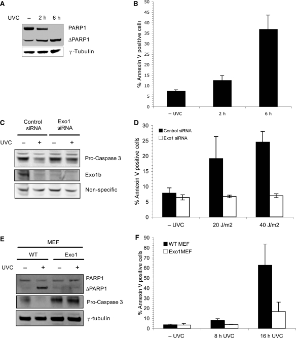
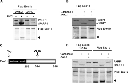
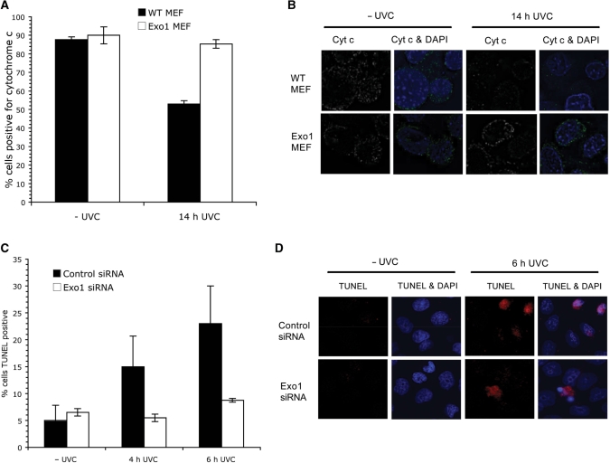
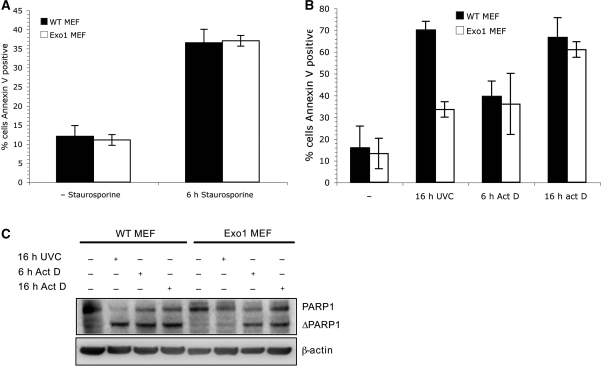
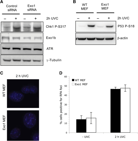
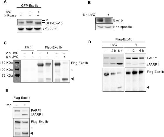
Apoptosis is essential for the maintenance of inherited genomic integrity. During DNA damage-induced apoptosis, mechanisms of cell survival, such as DNA repair are inactivated to allow cell death to proceed. Here, we describe a role for the mammalian DNA repair enzyme Exonuclease 1 (Exo1) in DNA damage-induced apoptosis. Depletion of Exo1 in human fibroblasts, or mouse embryonic fibroblasts led to a delay in DNA damage-induced apoptosis. Furthermore, we show that Exo1 acts upstream of caspase-3, DNA fragmentation and cytochrome c release. In addition, induction of apoptosis with DNA-damaging agents led to cleavage of both isoforms of Exo1. The cleavage of Exo1 was mapped to Asp514, and shown to be mediated by caspase-3. Expression of a caspase-3 cleavage site mutant form of Exo1, Asp514Ala, prevented formation of the previously observed fragment without any affect on the onset of apoptosis. We conclude that Exo1 has a role in the timely induction of apoptosis and that it is subsequently cleaved and degraded during apoptosis, potentially inhibiting DNA damage repair.
细胞凋亡对于维持遗传基因组的完整性至关重要。在DNA损伤诱导的细胞凋亡过程中,诸如DNA修复等细胞存活机制会失活,从而使细胞死亡得以进行。在此,我们描述了哺乳动物DNA修复酶核酸外切酶1(Exo1)在DNA损伤诱导的细胞凋亡中的作用。在人成纤维细胞或小鼠胚胎成纤维细胞中敲除Exo1会导致DNA损伤诱导的细胞凋亡延迟。此外,我们表明Exo1在半胱天冬酶-3、DNA片段化和细胞色素c释放的上游起作用。另外,用DNA损伤剂诱导细胞凋亡会导致Exo1的两种同工型均被切割。Exo1的切割位点定位于Asp514,并且显示由半胱天冬酶-3介导。Exo1的半胱天冬酶-3切割位点突变体形式Asp514Ala的表达可阻止先前观察到的片段的形成,而对细胞凋亡的起始没有任何影响。我们得出结论,Exo1在细胞凋亡的及时诱导中起作用,并且随后在细胞凋亡过程中被切割和降解,这可能会抑制DNA损伤修复。