• 文献检索
  • 文档翻译
  • 深度研究
  • 学术资讯
  • Suppr Zotero 插件Zotero 插件
  • 邀请有礼
  • 套餐&价格
  • 历史记录
应用&插件
Suppr Zotero 插件Zotero 插件浏览器插件Mac 客户端Windows 客户端微信小程序
定价
高级版会员购买积分包购买API积分包
服务
文献检索文档翻译深度研究API 文档MCP 服务
关于我们
关于 Suppr公司介绍联系我们用户协议隐私条款
关注我们

Suppr 超能文献

核心技术专利:CN118964589B侵权必究
粤ICP备2023148730 号-1Suppr @ 2026

文献检索

告别复杂PubMed语法,用中文像聊天一样搜索,搜遍4000万医学文献。AI智能推荐,让科研检索更轻松。

立即免费搜索

文件翻译

保留排版,准确专业,支持PDF/Word/PPT等文件格式,支持 12+语言互译。

免费翻译文档

深度研究

AI帮你快速写综述,25分钟生成高质量综述,智能提取关键信息,辅助科研写作。

立即免费体验

大鼠脑中T1和T2弛豫率(ΔR1和ΔR2)的时间变化与元素锰的组织清除率一致。

Temporal changes in the T1 and T2 relaxation rates (DeltaR1 and DeltaR2) in the rat brain are consistent with the tissue-clearance rates of elemental manganese.

作者信息

Chuang Kai-Hsiang, Koretsky Alan P, Sotak Christopher H

机构信息

Laboratory of Functional and Molecular Imaging, National Institute of Neurological Disorders and Stroke, National Institutes of Health, Bethesda, Maryland, USA.

出版信息

Magn Reson Med. 2009 Jun;61(6):1528-32. doi: 10.1002/mrm.21962.

DOI:10.1002/mrm.21962
PMID:19353652
原文链接:https://pmc.ncbi.nlm.nih.gov/articles/PMC2756245/
Abstract

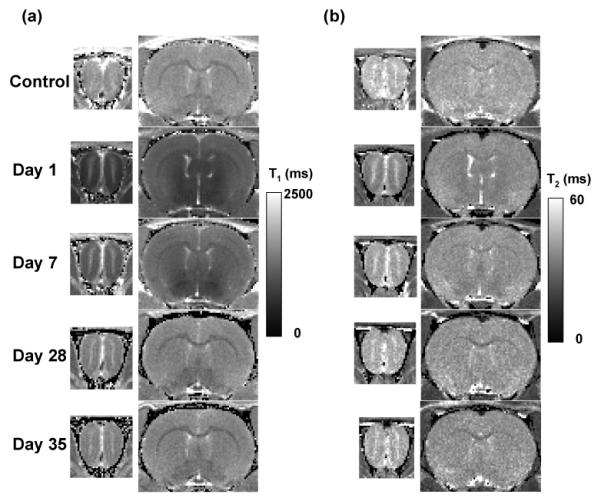
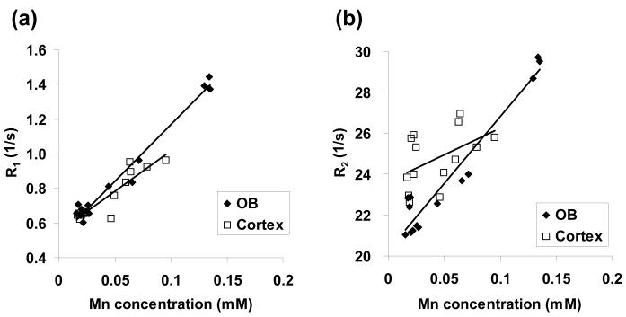
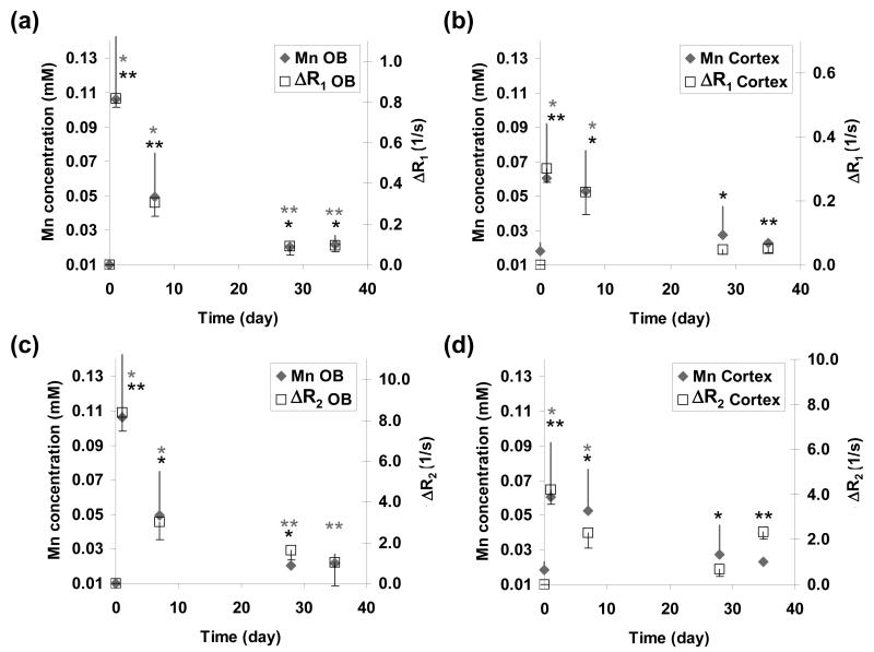
Temporal changes in the T(1) and T(2) relaxation rates (DeltaR(1) and DeltaR(2)) in rat olfactory bulb (OB) and cortex were compared with the absolute manganese (Mn) concentrations from the corresponding excised tissue samples. In vivo T(1) and T(2) relaxation times were measured before, and at 1, 7, 28, and 35 d after intravenous infusion of 176 mg/kg MnCl(2). The values of DeltaR(1), DeltaR(2), and absolute Mn concentration peaked at day 1 and then declined to near control levels after 28 to 35 d. The Mn bioelimination rate from the rat brain was significantly faster than that reported using radioisotope techniques. The R(1) and R(2) relaxation rates were linearly proportional to the underlying tissue Mn concentration and reflect the total absolute amount of Mn present in the tissue. The in vivo Mn r(1) and r(2) tissue relaxivities were comparable to the in vitro values for aqueous Mn(2+). These results demonstrate that loss of manganese-enhanced MRI (MEMRI) contrast after systemic Mn(2+) administration is due to elimination of Mn(2+) from the brain.

摘要

将大鼠嗅球(OB)和皮质中T(1)和T(2)弛豫率(ΔR(1)和ΔR(2))的时间变化与相应切除组织样本中的绝对锰(Mn)浓度进行了比较。在静脉注射176 mg/kg MnCl(2)之前以及之后的1、7、28和35天测量体内T(1)和T(2)弛豫时间。ΔR(1)、ΔR(2)和绝对Mn浓度的值在第1天达到峰值,然后在28至35天后降至接近对照水平。大鼠脑中Mn的生物消除率明显快于使用放射性同位素技术报道的速率。R(1)和R(2)弛豫率与基础组织Mn浓度呈线性比例,并反映组织中存在的Mn的总绝对量。体内Mn的r(1)和r(2)组织弛豫率与体外Mn(2+)水溶液的值相当。这些结果表明,全身给予Mn(2+)后锰增强磁共振成像(MEMRI)对比度的丧失是由于Mn(2+)从脑中消除所致。

https://cdn.ncbi.nlm.nih.gov/pmc/blobs/2f0d/2756245/6e95bfafd038/nihms-120394-f0003.jpg
https://cdn.ncbi.nlm.nih.gov/pmc/blobs/2f0d/2756245/06a4014df2fa/nihms-120394-f0001.jpg
https://cdn.ncbi.nlm.nih.gov/pmc/blobs/2f0d/2756245/9a89d8e4f283/nihms-120394-f0002.jpg
https://cdn.ncbi.nlm.nih.gov/pmc/blobs/2f0d/2756245/6e95bfafd038/nihms-120394-f0003.jpg
https://cdn.ncbi.nlm.nih.gov/pmc/blobs/2f0d/2756245/06a4014df2fa/nihms-120394-f0001.jpg
https://cdn.ncbi.nlm.nih.gov/pmc/blobs/2f0d/2756245/9a89d8e4f283/nihms-120394-f0002.jpg
https://cdn.ncbi.nlm.nih.gov/pmc/blobs/2f0d/2756245/6e95bfafd038/nihms-120394-f0003.jpg

相似文献

1
Temporal changes in the T1 and T2 relaxation rates (DeltaR1 and DeltaR2) in the rat brain are consistent with the tissue-clearance rates of elemental manganese.大鼠脑中T1和T2弛豫率(ΔR1和ΔR2)的时间变化与元素锰的组织清除率一致。
Magn Reson Med. 2009 Jun;61(6):1528-32. doi: 10.1002/mrm.21962.
2
Experimental protocol for activation-induced manganese-enhanced MRI (AIM-MRI) based on quantitative determination of Mn content in rat brain by fast T1 mapping.基于快速 T1 映射定量测定大鼠脑内 Mn 含量的激活诱导锰增强 MRI(AIM-MRI)的实验方案。
Magn Reson Med. 2009 Oct;62(4):1080-4. doi: 10.1002/mrm.22095.
3
Dose dependence and temporal evolution of the T1 relaxation time and MRI contrast in the rat brain after subcutaneous injection of manganese chloride.氯化锰皮下注射后大鼠脑 T1 弛豫时间和 MRI 对比的剂量依赖性和时变特征。
Magn Reson Med. 2012 Dec;68(6):1955-62. doi: 10.1002/mrm.24184. Epub 2012 Jan 31.
4
Manganese-enhanced magnetic resonance imaging (MEMRI) of rat brain after systemic administration of MnCl2: changes in T1 relaxation times during postnatal development.全身注射氯化锰后大鼠脑的锰增强磁共振成像(MEMRI):出生后发育过程中T1弛豫时间的变化
J Magn Reson Imaging. 2007 Jan;25(1):32-8. doi: 10.1002/jmri.20792.
5
Phase-based manganese enhanced MRI, a new methodology to enhance brain cytoarchitectural contrast and study manganese uptake.基于相位的锰增强磁共振成像,一种增强脑细结构对比度并研究锰摄取的新方法。
Magn Reson Med. 2014 Nov;72(5):1246-56. doi: 10.1002/mrm.25037. Epub 2013 Nov 20.
6
Manganese-enhanced MRI of the rat visual pathway: acute neural toxicity, contrast enhancement, axon resolution, axonal transport, and clearance of Mn(2+).大鼠视觉通路的锰增强磁共振成像:急性神经毒性、对比增强、轴突分辨率、轴突运输及锰离子清除
J Magn Reson Imaging. 2008 Oct;28(4):855-65. doi: 10.1002/jmri.21504.
7
Transcranial manganese delivery for neuronal tract tracing using MEMRI.经颅递送锰用于 MEMRI 示踪神经元束
Neuroimage. 2017 Aug 1;156:146-154. doi: 10.1016/j.neuroimage.2017.05.025. Epub 2017 May 13.
8
Infusion-based manganese-enhanced MRI: a new imaging technique to visualize the mouse brain.基于输注的锰增强 MRI:一种可视化小鼠大脑的新成像技术。
Brain Struct Funct. 2012 Jan;217(1):107-14. doi: 10.1007/s00429-011-0324-y. Epub 2011 May 20.
9
Quantitative activity-induced manganese-dependent MRI for characterizing cortical layers in the primary somatosensory cortex of the rat.定量活动诱导的锰依赖磁共振成像用于表征大鼠初级体感皮层的皮质层。
Brain Struct Funct. 2016 Mar;221(2):695-707. doi: 10.1007/s00429-014-0933-3. Epub 2014 Nov 1.
10
Direct CSF injection of MnCl(2) for dynamic manganese-enhanced MRI.经脑脊液直接注射氯化锰用于动态锰增强磁共振成像
Magn Reson Med. 2004 May;51(5):978-87. doi: 10.1002/mrm.20047.

引用本文的文献

1
From axonal transport to mitochondrial trafficking: What can we learn from Manganese-Enhanced MRI studies in mouse models of Alzheimers disease?从轴突运输到线粒体运输:在阿尔茨海默病小鼠模型的锰增强磁共振成像研究中我们能学到什么?
Curr Med Imaging Rev. 2011;7(1):16-27. doi: 10.2174/157340511794653478.
2
Genetic control of MRI contrast using the manganese transporter Zip14.利用锰转运蛋白 Zip14 进行 MRI 对比的遗传控制。
Magn Reson Med. 2024 Aug;92(2):820-835. doi: 10.1002/mrm.29993. Epub 2024 Apr 4.
3
In vivo MRI evaluation of anterograde manganese transport along the visual pathway following whole eye transplantation.

本文引用的文献

1
Manganese cell labeling of murine hepatocytes using manganese(III)-transferrin.使用锰(III)-转铁蛋白对小鼠肝细胞进行锰细胞标记
Contrast Media Mol Imaging. 2008 May-Jun;3(3):95-105. doi: 10.1002/cmmi.235.
2
Cerebrospinal fluid to brain transport of manganese in a non-human primate revealed by MRI.MRI揭示非人类灵长类动物中脑脊液向脑内转运锰的过程。
Brain Res. 2008 Mar 10;1198:160-70. doi: 10.1016/j.brainres.2007.12.065. Epub 2008 Jan 4.
3
Improved neuronal tract tracing using manganese enhanced magnetic resonance imaging with fast T(1) mapping.
全眼球移植后顺行锰转运沿视觉通路的活体 MRI 评估。
J Neurosci Methods. 2022 Apr 15;372:109534. doi: 10.1016/j.jneumeth.2022.109534. Epub 2022 Feb 22.
4
Chronic Intestinal Inflammation Suppresses Brain Activity by Inducing Neuroinflammation in Mice.慢性肠道炎症通过在小鼠中诱导神经炎症来抑制大脑活动。
Am J Pathol. 2022 Jan;192(1):72-86. doi: 10.1016/j.ajpath.2021.09.006. Epub 2021 Oct 5.
5
Applications of Manganese-Enhanced Magnetic Resonance Imaging in Ophthalmology and Visual Neuroscience.锰增强磁共振成像在眼科学和视觉神经科学中的应用。
Front Neural Circuits. 2019 May 14;13:35. doi: 10.3389/fncir.2019.00035. eCollection 2019.
6
Activity-induced MEMRI cannot detect functional brain anomalies in the APPxPS1-Ki mouse model of Alzheimer's disease.活动诱导的 MEMRI 无法检测阿尔茨海默病 APPxPS1-Ki 小鼠模型中的功能性脑异常。
Sci Rep. 2019 Feb 4;9(1):1140. doi: 10.1038/s41598-018-37980-y.
7
Plasticity changes in forebrain activity and functional connectivity during neuropathic pain development in rats with sciatic spared nerve injury.在坐骨神经部分损伤大鼠神经病理性疼痛发展过程中,前脑活动和功能连接的可塑性变化。
Mol Brain. 2018 Oct 1;11(1):55. doi: 10.1186/s13041-018-0398-z.
8
Tinnitus and temporary hearing loss result in differential noise-induced spatial reorganization of brain activity.耳鸣和暂时性听力损失导致了不同的噪声诱导的大脑活动空间重组。
Brain Struct Funct. 2018 Jun;223(5):2343-2360. doi: 10.1007/s00429-018-1635-z. Epub 2018 Feb 27.
9
Development of manganese-enhanced magnetic resonance imaging of the rostral ventrolateral medulla of conscious rats: Importance of normalization and comparison with other regions of interest.清醒大鼠延髓头端腹外侧区锰增强磁共振成像的发展:标准化的重要性及与其他感兴趣区域的比较
NMR Biomed. 2018 Mar;31(3). doi: 10.1002/nbm.3887. Epub 2018 Jan 12.
10
Thalamic GABA levels and occupational manganese neurotoxicity: Association with exposure levels and brain MRI.丘脑 GABA 水平与职业性锰神经毒性:与暴露水平和脑 MRI 的关联。
Neurotoxicology. 2018 Jan;64:30-42. doi: 10.1016/j.neuro.2017.08.013. Epub 2017 Sep 2.
利用快速T(1) 映射的锰增强磁共振成像改进神经元束示踪
Magn Reson Med. 2006 Mar;55(3):604-11. doi: 10.1002/mrm.20797.
4
Manganese-enhanced magnetic resonance imaging (MEMRI): methodological and practical considerations.锰增强磁共振成像(MEMRI):方法学与实际考量
NMR Biomed. 2004 Dec;17(8):532-43. doi: 10.1002/nbm.945.
5
Intracellular manganese ions provide strong T1 relaxation in rat myocardium.细胞内锰离子在大鼠心肌中提供强烈的T1弛豫。
Magn Reson Med. 2004 Sep;52(3):506-14. doi: 10.1002/mrm.20199.
6
In vivo detection of neuroarchitecture in the rodent brain using manganese-enhanced MRI.利用锰增强磁共振成像在啮齿动物大脑中进行神经结构的体内检测。
Neuroimage. 2004 Jul;22(3):1046-59. doi: 10.1016/j.neuroimage.2004.03.031.
7
Direct CSF injection of MnCl(2) for dynamic manganese-enhanced MRI.经脑脊液直接注射氯化锰用于动态锰增强磁共振成像
Magn Reson Med. 2004 May;51(5):978-87. doi: 10.1002/mrm.20047.
8
Manganese ions as intracellular contrast agents: proton relaxation and calcium interactions in rat myocardium.锰离子作为细胞内造影剂:大鼠心肌中的质子弛豫和钙相互作用
NMR Biomed. 2003 Apr;16(2):82-95. doi: 10.1002/nbm.817.
9
Manganese neurotoxicity: an update of pathophysiologic mechanisms.锰神经毒性:病理生理机制的最新进展
Metab Brain Dis. 2002 Dec;17(4):375-87. doi: 10.1023/a:1021970120965.
10
Manganese action in brain function.锰在脑功能中的作用。
Brain Res Brain Res Rev. 2003 Jan;41(1):79-87. doi: 10.1016/s0165-0173(02)00234-5.