Huereca Daniel J, Bakoulas Konstandinos A, Ghoddoussi Farhad, Berkowitz Bruce A, Holt Avril Genene, Mueller Patrick J
Department of Pharmacology, Wayne State University School of Medicine, Detroit, MI, USA.
Department of Physiology, Wayne State University School of Medicine, Detroit, MI, USA.
NMR Biomed. 2018 Mar;31(3). doi: 10.1002/nbm.3887. Epub 2018 Jan 12.
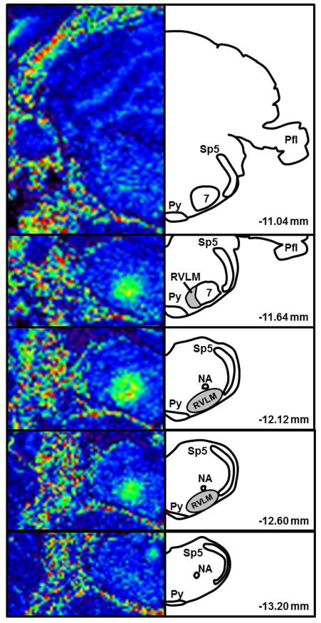
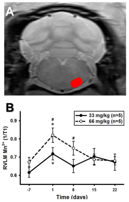
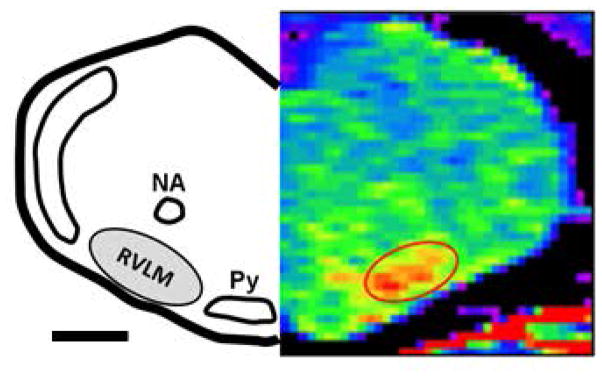
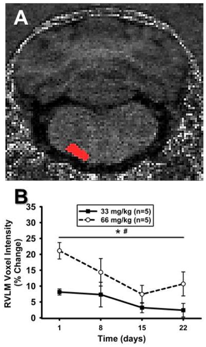
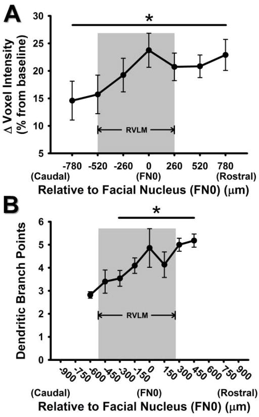
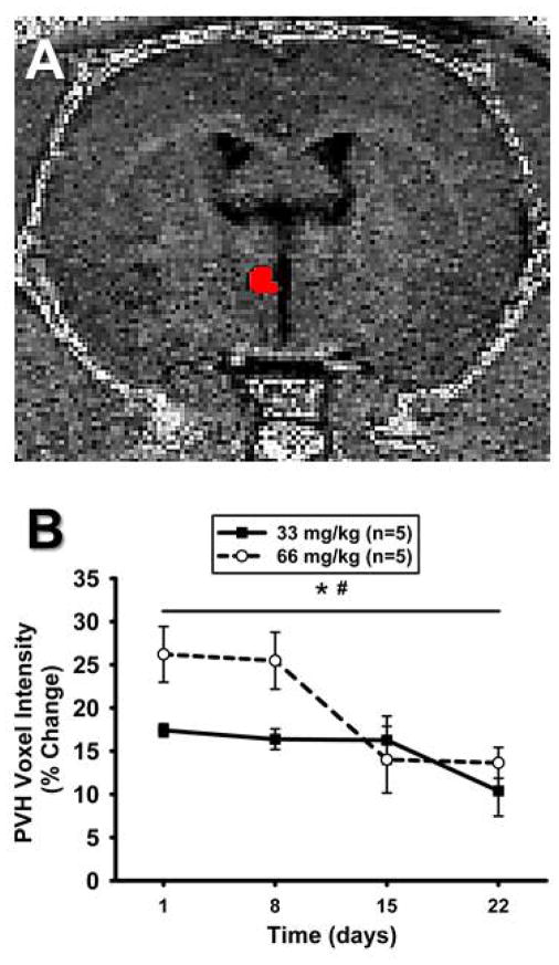
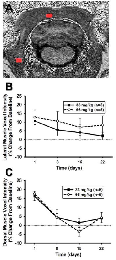
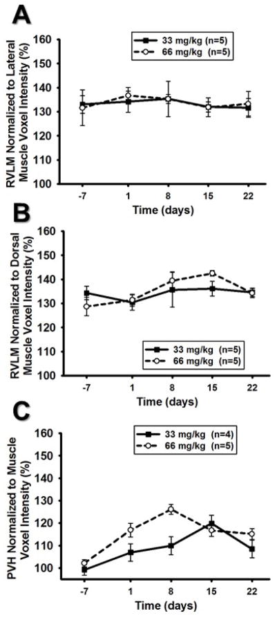
Spinally projecting neurons in the rostral ventrolateral medulla (RVLM) are believed to contribute to pathophysiological alterations in sympathetic nerve activity and the development of cardiovascular disease. The ability to identify changes in the activity of RVLM neurons in conscious animals and humans, especially longitudinally, would represent a clinically important advancement in our understanding of the contribution of the RVLM to cardiovascular disease. To this end, we describe the initial development of manganese-enhanced magnetic resonance imaging (MEMRI) for the rat RVLM. Manganese (Mn ) has been used to estimate in vivo neuronal activity in other brain regions because of both its paramagnetic properties and its entry into and accumulation in active neurons. In this initial study, our three goals were as follows: (1) to validate that Mn enhancement occurs in functionally and anatomically localized images of the rat RVLM; (2) to quantify the dose and time course dependence of Mn enhancement in the RVLM after one systemic injection in conscious rats (66 or 33 mg/kg, intraperitoneally); and (3) to compare Mn enhancement in the RVLM with other regions to determine an appropriate method of normalization of T -weighted images. In our proof-of-concept and proof-of-principle studies, Mn was identified by MRI in the rat RVLM after direct microinjection or via retrograde transport following spinal cord injections, respectively. Systemic injections in conscious rats produced significant Mn enhancement at 24 h (p < 0.05). Injections of 66 mg/kg produced greater enhancement than 33 mg/kg in the RVLM and paraventricular nucleus of the hypothalamus (p < 0.05 for both), but only when normalized to baseline scans without Mn injection. Consistent with findings from our previous functional and anatomical studies demonstrating subregional neuroplasticity, Mn enhancement was higher in the rostral regions of the RVLM (p < 0.05). Together with important technical considerations, our studies support the development of MEMRI as a potential method to examine RVLM activity over time in conscious animal subjects.
延髓头端腹外侧区(RVLM)中向脊髓投射的神经元被认为与交感神经活动的病理生理改变以及心血管疾病的发展有关。能够识别清醒动物和人类中RVLM神经元活动的变化,尤其是纵向变化,将是我们在理解RVLM对心血管疾病的作用方面取得的一项具有临床重要意义的进展。为此,我们描述了用于大鼠RVLM的锰增强磁共振成像(MEMRI)的初步开发。锰(Mn)因其顺磁性特性以及进入并积聚在活跃神经元中,已被用于估计其他脑区的体内神经元活动。在这项初步研究中,我们的三个目标如下:(1)验证在大鼠RVLM的功能和解剖定位图像中是否出现锰增强;(2)在清醒大鼠(腹腔注射66或33 mg/kg)单次全身注射后,量化RVLM中锰增强的剂量和时间进程依赖性;(3)比较RVLM与其他区域的锰增强情况,以确定T加权图像的合适归一化方法。在我们的概念验证和原理验证研究中,分别通过直接微量注射或脊髓注射后的逆行转运,在大鼠RVLM中通过MRI识别出了锰。清醒大鼠的全身注射在24小时时产生了显著的锰增强(p < 0.05)。66 mg/kg的注射在RVLM和下丘脑室旁核中产生的增强比33 mg/kg更大(两者均p < 0.05),但只有在归一化至未注射锰的基线扫描时才如此。与我们之前证明亚区域神经可塑性的功能和解剖学研究结果一致,RVLM头端区域的锰增强更高(p < 0.05)。连同重要的技术考虑因素,我们的研究支持将MEMRI开发为一种在清醒动物受试者中随时间检查RVLM活动的潜在方法。