Schallek Jesse, Kardon Randy, Kwon Young, Abramoff Michael, Soliz Peter, Ts'o Daniel
Department of Neurosurgery, SUNY Upstate Medical University, Syracuse, New York 13210, USA.
Invest Ophthalmol Vis Sci. 2009 Oct;50(10):4873-80. doi: 10.1167/iovs.08-3291. Epub 2009 May 6.
To elucidate the anatomic origins of stimulus-evoked intrinsic optical signals in the mammalian retina by using selective pharmacologic blockade of specific retinal layers.
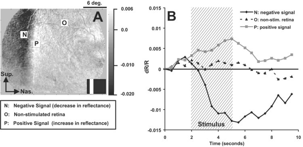
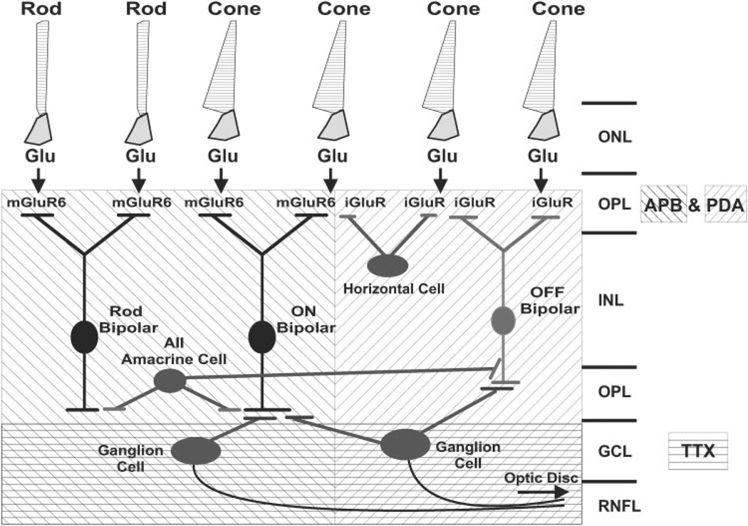
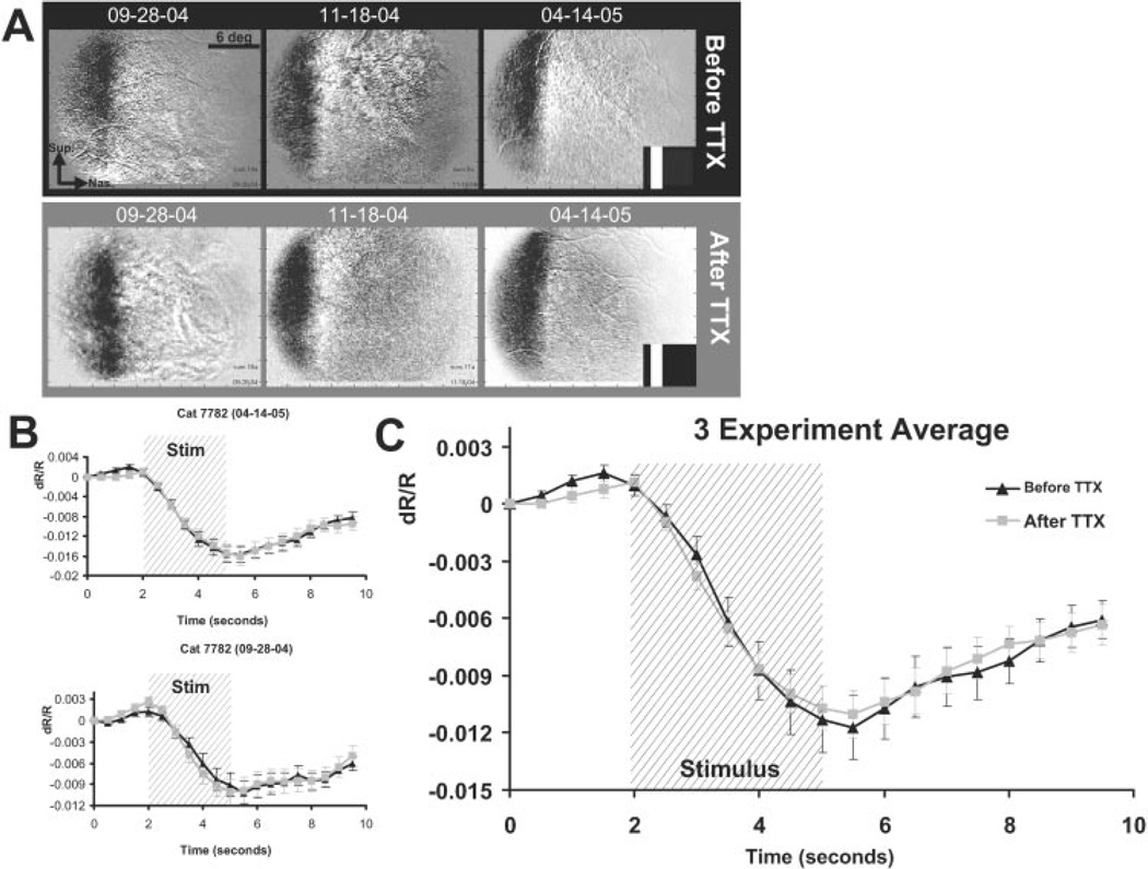
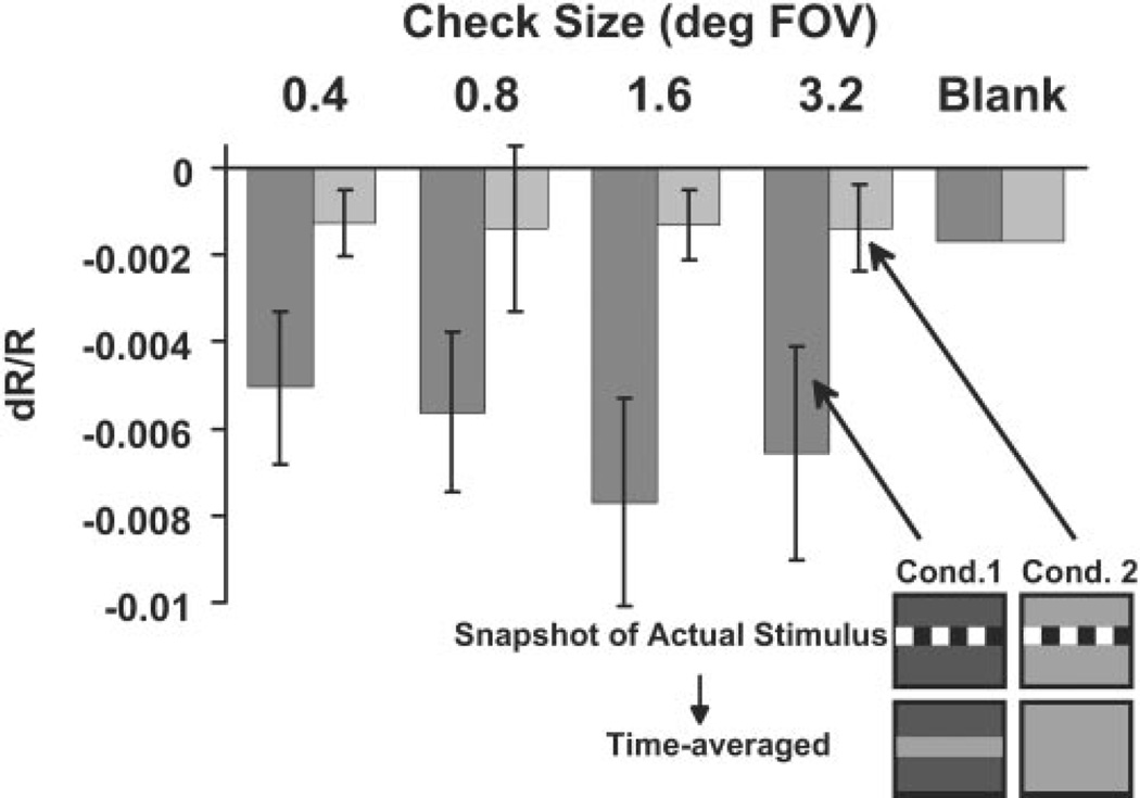
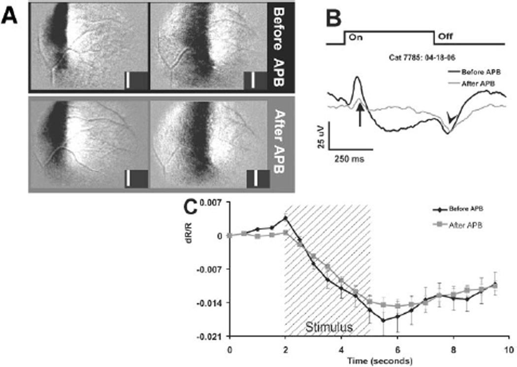
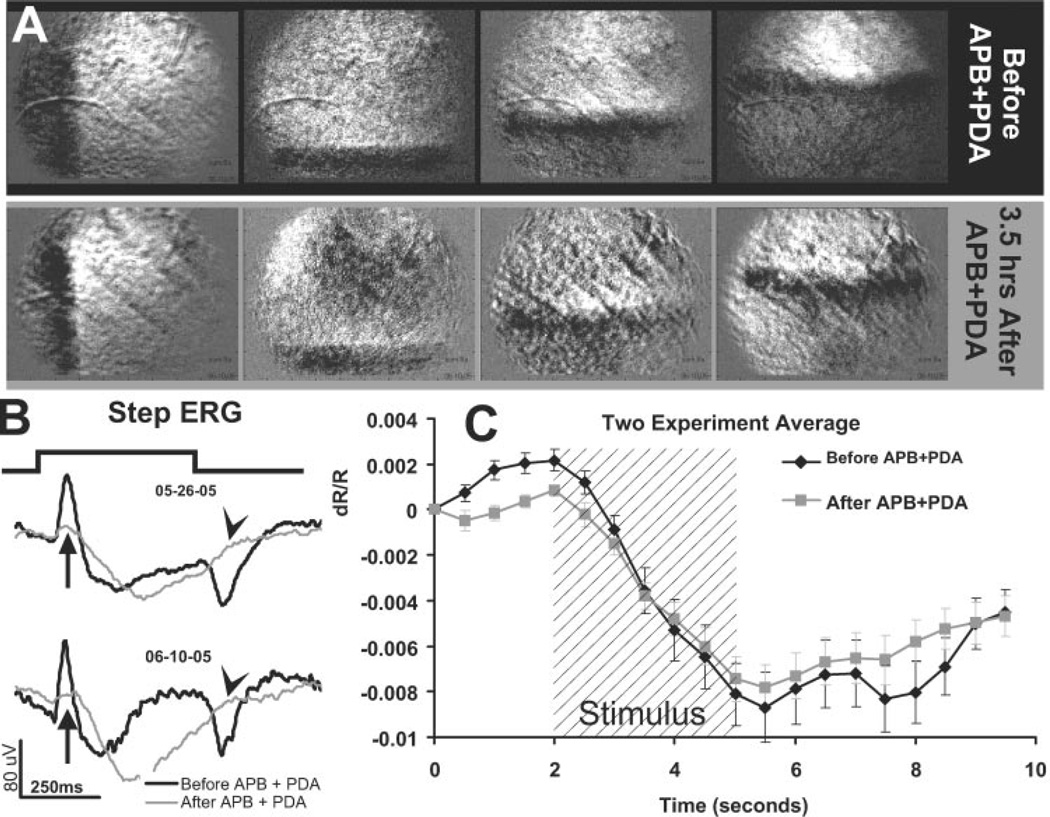
Four adult cats were used to investigate the stimulus-evoked intrinsic signals. The retinas were visually stimulated with a liquid crystal display (LCD) integrated into a modified fundus camera. The evoked signals in the near infrared (NIR) were recorded with a digital camera to image the changes in the optical reflectance of the retinas. Variants of the electroretinogram (pattern ERG and long-pulse ERG) were also recorded as additional measures of retinal function. Specific retinal layers were inactivated via intravitreal injections of the voltage-gated sodium channel blocker, tetrodotoxin (TTX), the metabotropic glutamate receptor (mGluR6) agonist, 2-amino-4-phosphonobutyric acid (APB), and/or the ionotropic glutamate receptor antagonist cis-2,3 piperidinedicarboxylic acid (PDA). The stimulus-evoked intrinsic signals were imaged before and after drug injection.
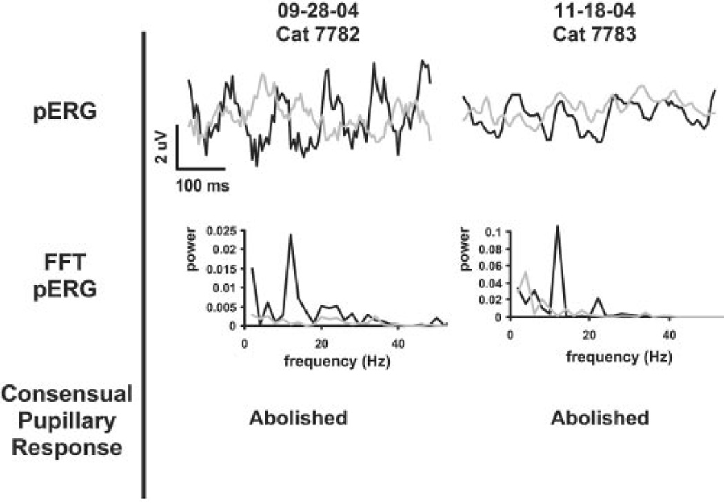
ERG recordings and tests of the consensual pupillary response confirmed the effectiveness of each drug. Yet despite the pharmacologic blockade of the inner retina (TTX) and postreceptoral retinal circuitry (APB and PDA), the stimulus-evoked intrinsic signals remained essentially unaltered from preinjection conditions. Similarly, the time course of the signal did not appreciably shift in time or shape.
The findings demonstrate that stimulus-evoked intrinsic signals persist after injection of APB, PDA, and TTX, drugs that work to suppress inner and postreceptoral retinal circuitry. The persistence of the intrinsic signals after administration of these drugs indicates that the dominant intrinsic signals are likely to arise from the outer retina.
通过对特定视网膜层进行选择性药物阻断,阐明哺乳动物视网膜中刺激诱发的内在光学信号的解剖学起源。
使用4只成年猫来研究刺激诱发的内在信号。视网膜通过集成在改良眼底相机中的液晶显示器(LCD)进行视觉刺激。用数码相机记录近红外(NIR)中的诱发信号,以成像视网膜光学反射率的变化。视网膜电图的变体(图形视网膜电图和长脉冲视网膜电图)也作为视网膜功能的额外测量指标进行记录。通过玻璃体内注射电压门控钠通道阻滞剂河豚毒素(TTX)、代谢型谷氨酸受体(mGluR6)激动剂2-氨基-4-磷酸丁酸(APB)和/或离子型谷氨酸受体拮抗剂顺式-2,3-哌啶二羧酸(PDA)使特定视网膜层失活。在药物注射前后对刺激诱发的内在信号进行成像。
视网膜电图记录和对交感瞳孔反应的测试证实了每种药物的有效性。然而,尽管对内视网膜(TTX)以及感受器后视网膜回路(APB和PDA)进行了药物阻断,但刺激诱发的内在信号与注射前相比基本未变。同样,信号的时间进程在时间或形状上也没有明显变化。
研究结果表明,在注射APB、PDA和TTX(这些药物可抑制内视网膜和感受器后视网膜回路)后,刺激诱发的内在信号仍然存在。施用这些药物后内在信号的持续存在表明,主要的内在信号可能源自外视网膜。