Matheus Ricardo Alves, Ramos-Perez Flávia Maria de Moraes, Menezes Alynne Vieira, Ambrosano Gláucia Maria Bovi, Haiter-Neto Francisco, Bóscolo Frab Norberto, de Almeida Solange Maria
Dental School of Piracicaba, State University of Campinas, Piracicaba, SP, Brazil.
J Appl Oral Sci. 2009 May-Jun;17(3):204-8. doi: 10.1590/s1678-77572009000300014.
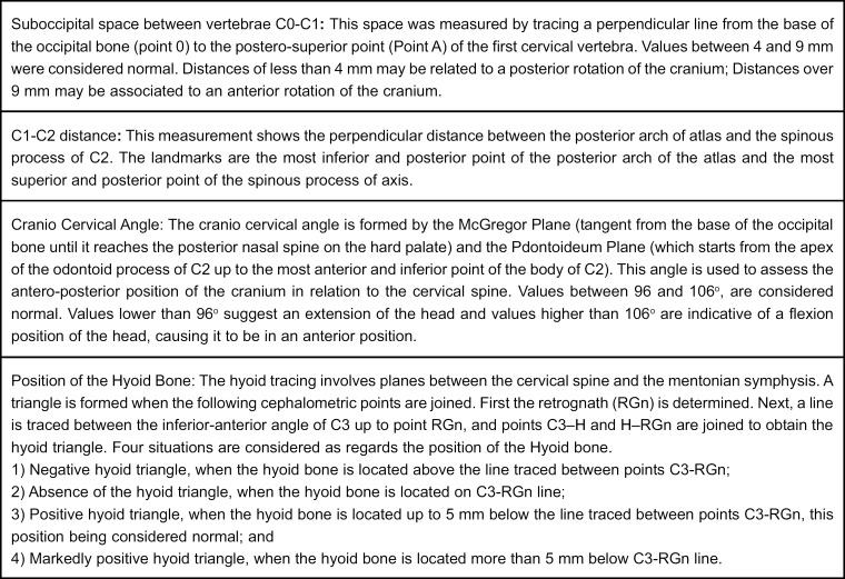
This study aimed to evaluate the possibility of any correlation between disc displacement and parameters used for evaluation of skull positioning in relation to the cervical spine: craniocervical angle, suboccipital space between C0-C1, cervical curvature and position of the hyoid bone in individuals with and without symptoms of temporomandibular dysfunction.
The patients were evaluated following the guidelines set forth by RDC/TMD. Evaluation was performed by magnetic resonance imaging for establishment of disc positioning in the temporomandibular joints (TMJs) of 30 volunteer patients without temporomandibular dysfunction symptoms and 30 patients with symptoms. Evaluation of skull positioning in relation to the cervical spine was performed on lateral cephalograms achieved with the individual in natural head position. Data were submitted to statistical analysis by Fisher's exact test at 5% significance level. To measure the degree of reproducibility/agreements between surveys, the kappa (K) statistics was used.
Significant differences were observed between C0-C1 measurement for both symptomatic (p=0.04) and asymptomatic (p=0.02). No statistical differences were observed regarding craniocervical angle, C1-C2 and hyoid bone position in relation to the TMJs with and without disc displacement. Although statistically significant difference was found in the C0-C1 space, no association between these and internal temporomandibular joint disorder can be considered.
Based on the results observed in this study, no direct relationship could be determined between the presence of disc displacement and the variables assessed.
本研究旨在评估在有和没有颞下颌关节紊乱症状的个体中,椎间盘移位与用于评估颅骨相对于颈椎位置的参数之间是否存在相关性,这些参数包括颅颈角、C0-C1之间的枕下间隙、颈椎曲度以及舌骨位置。
按照RDC/TMD制定的指南对患者进行评估。通过磁共振成像对30名无颞下颌关节紊乱症状的志愿者患者和30名有症状的患者的颞下颌关节(TMJ)中的椎间盘位置进行评估。在个体处于自然头位时获得的头颅侧位片上评估颅骨相对于颈椎的位置。数据采用Fisher精确检验进行统计学分析,显著性水平为5%。为了测量调查之间的可重复性/一致性程度,使用kappa(K)统计量。
在有症状组(p=0.04)和无症状组(p=0.02)的C0-C1测量值之间均观察到显著差异。在有无椎间盘移位的情况下,关于颅颈角、C1-C2和舌骨相对于TMJ的位置未观察到统计学差异。虽然在C0-C1间隙中发现了统计学上的显著差异,但不能认为这些与颞下颌关节内部紊乱之间存在关联。
基于本研究观察到的结果,无法确定椎间盘移位的存在与所评估变量之间的直接关系。