• 文献检索
  • 文档翻译
  • 深度研究
  • 学术资讯
  • Suppr Zotero 插件Zotero 插件
  • 邀请有礼
  • 套餐&价格
  • 历史记录
应用&插件
Suppr Zotero 插件Zotero 插件浏览器插件Mac 客户端Windows 客户端微信小程序
定价
高级版会员购买积分包购买API积分包
服务
文献检索文档翻译深度研究API 文档MCP 服务
关于我们
关于 Suppr公司介绍联系我们用户协议隐私条款
关注我们

Suppr 超能文献

核心技术专利:CN118964589B侵权必究
粤ICP备2023148730 号-1Suppr @ 2026

文献检索

告别复杂PubMed语法,用中文像聊天一样搜索,搜遍4000万医学文献。AI智能推荐,让科研检索更轻松。

立即免费搜索

文件翻译

保留排版,准确专业,支持PDF/Word/PPT等文件格式,支持 12+语言互译。

免费翻译文档

深度研究

AI帮你快速写综述,25分钟生成高质量综述,智能提取关键信息,辅助科研写作。

立即免费体验

磁共振方法对肝脏的参数探索。

Parametric exploration of the liver by magnetic resonance methods.

机构信息

Radiology, University Medical Center Groningen and University of Groningen, Hanzeplein 1, 9713 GZ, Groningen, The Netherlands.

出版信息

Eur Radiol. 2009 Nov;19(11):2594-607. doi: 10.1007/s00330-009-1470-y. Epub 2009 Jun 6.

DOI:10.1007/s00330-009-1470-y
PMID:19504103
原文链接:https://pmc.ncbi.nlm.nih.gov/articles/PMC2762052/
Abstract

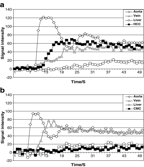
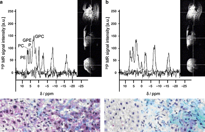
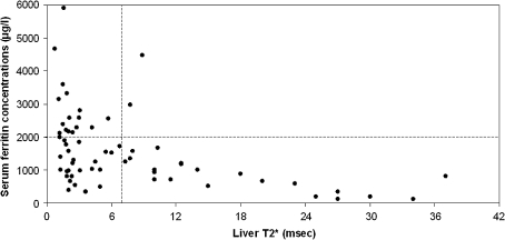
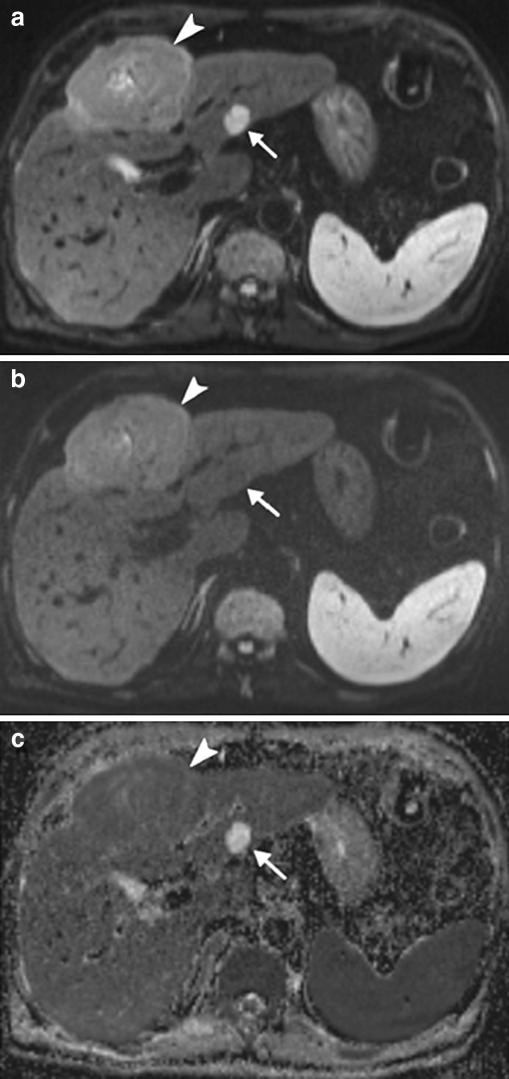
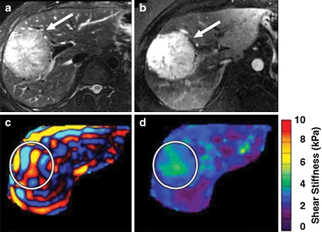
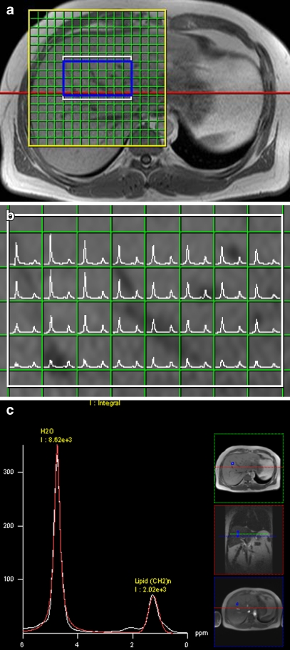
MRI, as a completely noninvasive technique, can provide quantitative assessment of perfusion, diffusion, viscoelasticity and metabolism, yielding diverse information about liver function. Furthermore, pathological accumulations of iron and lipids can be quantified. Perfusion MRI with various contrast agents is commonly used for the detection and characterization of focal liver disease and the quantification of blood flow parameters. An extended new application is the evaluation of the therapeutic effect of antiangiogenic drugs on liver tumours. Novel, but already widespread, is a histologically validated relaxometry method using five gradient echo sequences for quantifying liver iron content elevation, a measure of inflammation, liver disease and cancer. Because of the high perfusion fraction in the liver, the apparent diffusion coefficients strongly depend on the gradient factors used in diffusion-weighted MRI. While complicating analysis, this offers the opportunity to study perfusion without contrast injection. Another novel method, MR elastography, has already been established as the only technique able to stage fibrosis or diagnose mild disease. Liver fat content is accurately determined with multivoxel MR spectroscopy (MRS) or by faster MRI methods that are, despite their widespread use, prone to systematic error. Focal liver disease characterisation will be of great benefit once multivoxel methods with fat suppression are implemented in proton MRS, in particular on high-field MR systems providing gains in signal-to-noise ratio and spectral resolution.

摘要

MRI 作为一种完全无创的技术,可以提供灌注、扩散、粘弹性和代谢的定量评估,提供有关肝功能的多种信息。此外,还可以定量检测铁和脂质的病理性堆积。各种对比剂的灌注 MRI 常用于检测和描述局灶性肝病,并量化血流参数。一个扩展的新应用是评估抗血管生成药物对肝肿瘤的治疗效果。一种新的但已经广泛应用的方法是使用五个梯度回波序列进行组织学验证的弛豫率测量法,用于定量肝铁含量升高,这是炎症、肝病和癌症的一个指标。由于肝脏的灌注分数较高,表观扩散系数强烈依赖于扩散加权 MRI 中使用的梯度因子。虽然这使得分析变得复杂,但它提供了无需对比注射即可研究灌注的机会。另一种新方法,磁共振弹性成像,已经成为唯一能够分期纤维化或诊断轻度疾病的技术。多体素磁共振波谱(MRS)或更快的 MRI 方法可以准确确定肝内脂肪含量,尽管这些方法得到了广泛应用,但容易出现系统误差。一旦在质子 MRS 中实施带有脂肪抑制的多体素方法,特别是在提供信噪比和光谱分辨率增益的高场 MRI 系统上,局灶性肝病的特征描述将非常有益。

https://cdn.ncbi.nlm.nih.gov/pmc/blobs/4aa7/2762052/f95b883e1748/330_2009_1470_Fig6_HTML.jpg
https://cdn.ncbi.nlm.nih.gov/pmc/blobs/4aa7/2762052/f185144315f7/330_2009_1470_Fig1_HTML.jpg
https://cdn.ncbi.nlm.nih.gov/pmc/blobs/4aa7/2762052/9193ea26373e/330_2009_1470_Fig2_HTML.jpg
https://cdn.ncbi.nlm.nih.gov/pmc/blobs/4aa7/2762052/90b3de9929b7/330_2009_1470_Fig3_HTML.jpg
https://cdn.ncbi.nlm.nih.gov/pmc/blobs/4aa7/2762052/e3bf330d0c4d/330_2009_1470_Fig4_HTML.jpg
https://cdn.ncbi.nlm.nih.gov/pmc/blobs/4aa7/2762052/8f7ebf54328c/330_2009_1470_Fig5_HTML.jpg
https://cdn.ncbi.nlm.nih.gov/pmc/blobs/4aa7/2762052/f95b883e1748/330_2009_1470_Fig6_HTML.jpg

相似文献

1
Parametric exploration of the liver by magnetic resonance methods.磁共振方法对肝脏的参数探索。
Eur Radiol. 2009 Nov;19(11):2594-607. doi: 10.1007/s00330-009-1470-y. Epub 2009 Jun 6.
2
Liver fibrosis: Review of current imaging and MRI quantification techniques.肝纤维化:当前影像学及磁共振成像定量技术综述
J Magn Reson Imaging. 2017 May;45(5):1276-1295. doi: 10.1002/jmri.25550. Epub 2016 Dec 16.
3
Liver Fibrosis Quantification by Magnetic Resonance Imaging.通过磁共振成像进行肝纤维化定量分析
Top Magn Reson Imaging. 2017 Dec;26(6):229-241. doi: 10.1097/RMR.0000000000000149.
4
Updates on Quantitative MRI of Diffuse Liver Disease: A Narrative Review.漫谈肝脏弥散性疾病的定量 MRI 研究进展
Biomed Res Int. 2022 Dec 28;2022:1147111. doi: 10.1155/2022/1147111. eCollection 2022.
5
New imaging techniques for liver diseases.肝脏疾病的新型影像学技术。
J Hepatol. 2015 Mar;62(3):690-700. doi: 10.1016/j.jhep.2014.10.014. Epub 2014 Oct 17.
6
Prospective Evaluation of Virtual MR Elastography With Diffusion-Weighted Imaging in Subjects With Nonalcoholic Fatty Liver Disease.前瞻性评估弥散加权成像虚拟磁共振弹性成像在非酒精性脂肪性肝病患者中的应用。
J Magn Reson Imaging. 2022 Nov;56(5):1448-1456. doi: 10.1002/jmri.28154. Epub 2022 Mar 14.
7
Is MR spectroscopy really the best MR-based method for the evaluation of fatty liver in diabetic patients in clinical practice?在临床实践中,磁共振波谱法真的是评估糖尿病患者脂肪肝的最佳基于磁共振的方法吗?
PLoS One. 2014 Nov 26;9(11):e112574. doi: 10.1371/journal.pone.0112574. eCollection 2014.
8
[Magnetic resonance imaging for quantifying hepatis steatosis and hepatic fibrosis].[用于量化肝脂肪变性和肝纤维化的磁共振成像]
Bull Mem Acad R Med Belg. 2009;164(1-2):44-50; discussion 51-2.
9
Contribution of diffusion-weighted magnetic resonance imaging in the characterization of hepatocellular carcinomas and dysplastic nodules in cirrhotic liver.扩散加权磁共振成像在肝硬化肝脏中肝细胞癌和发育异常结节特征描述中的作用
J Comput Assist Tomogr. 2010 Jul;34(4):506-12. doi: 10.1097/RCT.0b013e3181da3671.
10
A comparison of liver fat content as determined by magnetic resonance imaging-proton density fat fraction and MRS versus liver histology in non-alcoholic fatty liver disease.磁共振成像质子密度脂肪分数和磁共振波谱测定的肝脏脂肪含量与非酒精性脂肪性肝病肝脏组织学的比较
Acta Radiol. 2016 Mar;57(3):271-8. doi: 10.1177/0284185115580488. Epub 2015 Apr 8.

引用本文的文献

1
Chinese tree shrews as a primate experimental animal eligible for the study of alcoholic liver disease: characterization and confirmation by MRI.中国树鼩作为一种适合研究酒精性肝病的灵长类实验动物:磁共振成像的特征和确认。
Exp Anim. 2020 Jan 29;69(1):110-118. doi: 10.1538/expanim.19-0073. Epub 2019 Sep 26.
2
Evaluation of liver fibrosis with a monoexponential model of intravoxel incoherent motion magnetic resonance imaging.基于体素内不相干运动磁共振成像单指数模型评估肝纤维化
Oncotarget. 2018 May 15;9(37):24619-24626. doi: 10.18632/oncotarget.24758.
3
Protective Effect of Yang Mi Ryung® Extract on Noise-Induced Hearing Loss in Mice.

本文引用的文献

1
RETRACTED ARTICLE: Diagnosis of rejection after liver transplantation: use of phosphorus-31 magnetic resonance spectroscopy ((31)P-MRS).撤回文章:肝移植术后排斥反应的诊断:磷-31磁共振波谱法(³¹P-MRS)的应用
Abdom Imaging. 2012 Oct;37(5):788-94. doi: 10.1007/s00261-008-9451-1.
2
Unresectable hepatocellular carcinoma: serial early vascular and cellular changes after transarterial chemoembolization as detected with MR imaging.不可切除肝细胞癌:经动脉化疗栓塞术后早期血管及细胞的系列变化——磁共振成像检测结果
Radiology. 2009 Feb;250(2):466-73. doi: 10.1148/radiol.2502072222.
3
Imaging of hepatocellular carcinoma: qualitative and quantitative analysis of postvascular phase contrast-enhanced ultrasonography with sonazoid. Comparison with superparamagnetic iron oxide magnetic resonance images.
杨米灵®提取物对小鼠噪声性听力损失的保护作用。
Evid Based Complement Alternat Med. 2017;2017:9814836. doi: 10.1155/2017/9814836. Epub 2017 Nov 15.
4
The Clinical Implications of Fatty Pancreas: A Concise Review.胰腺脂肪化的临床意义:简要综述
Dig Dis Sci. 2017 Oct;62(10):2658-2667. doi: 10.1007/s10620-017-4700-1. Epub 2017 Aug 8.
5
Pancreatic fat and β-cell function in overweight/obese children with nonalcoholic fatty liver disease.非酒精性脂肪性肝病超重/肥胖儿童的胰腺脂肪与β细胞功能
World J Gastroenterol. 2015 Apr 21;21(15):4688-95. doi: 10.3748/wjg.v21.i15.4688.
6
3.0 T proton magnetic resonance spectroscopy of the liver: quantification of choline.肝脏 3.0T 质子磁共振波谱:胆碱定量分析。
World J Gastroenterol. 2013 Mar 7;19(9):1472-7. doi: 10.3748/wjg.v19.i9.1472.
7
Molecular MRI of liver fibrosis by a peptide-targeted contrast agent in an experimental mouse model.实验性小鼠模型中肽靶向对比剂的肝纤维化分子 MRI。
Invest Radiol. 2013 Jan;48(1):46-54. doi: 10.1097/RLI.0b013e3182749c0b.
8
Cross-sectional investigation of correlation between hepatic steatosis and IVIM perfusion on MR imaging.磁共振成像中肝脂肪变性与 IVIM 灌注相关性的横断面研究。
Magn Reson Imaging. 2012 May;30(4):572-8. doi: 10.1016/j.mri.2011.12.013. Epub 2012 Jan 27.
9
MR Imaging of activated hepatic stellate cells in liver injured by CCl4 of rats with integrin-targeted ultrasmall superparamagnetic iron oxide.整合素靶向超顺磁性氧化铁致肝损伤大鼠肝星状细胞激活的磁共振成像
Eur Radiol. 2011 May;21(5):1016-25. doi: 10.1007/s00330-010-1988-z. Epub 2010 Oct 25.
10
Assessment of relevant hepatic steatosis in obese adolescents by rapid fat-selective GRE imaging with spatial-spectral excitation: a quantitative comparison with spectroscopic findings.采用快速选择性 GRE 成像技术评估肥胖青少年的相关肝脂肪变:与光谱学发现的定量比较。
Eur Radiol. 2011 Apr;21(4):816-22. doi: 10.1007/s00330-010-1975-4. Epub 2010 Oct 3.
肝细胞癌的成像:使用声诺维进行血管后期对比增强超声检查的定性和定量分析。与超顺磁性氧化铁磁共振图像的比较。
Oncology. 2008;75 Suppl 1:48-54. doi: 10.1159/000173424. Epub 2008 Dec 17.
4
Monitoring of liver glycogen synthesis in diabetic patients using carbon-13 MR spectroscopy.应用碳-13 MR 波谱监测糖尿病患者肝糖原合成。
Eur J Radiol. 2010 Feb;73(2):300-4. doi: 10.1016/j.ejrad.2008.10.019. Epub 2008 Dec 5.
5
Can imaging modalities diagnose and stage hepatic fibrosis and cirrhosis accurately?成像模态能否准确诊断肝纤维化和肝硬化并进行分期?
J Hepatol. 2009 Jan;50(1):17-35. doi: 10.1016/j.jhep.2008.10.016. Epub 2008 Nov 8.
6
Multiecho MR imaging and proton MR spectroscopy for liver fat quantification.用于肝脏脂肪定量的多回波磁共振成像和质子磁共振波谱分析
Radiology. 2008 Dec;249(3):1081. doi: 10.1148/radiol.2493081034.
7
Liver cirrhosis: intravoxel incoherent motion MR imaging--pilot study.肝硬化:体素内不相干运动磁共振成像——初步研究。
Radiology. 2008 Dec;249(3):891-9. doi: 10.1148/radiol.2493080080.
8
Comparison and reproducibility of ADC measurements in breathhold, respiratory triggered, and free-breathing diffusion-weighted MR imaging of the liver.屏气、呼吸触发和自由呼吸肝脏扩散加权磁共振成像中表观扩散系数(ADC)测量值的比较及可重复性
J Magn Reson Imaging. 2008 Nov;28(5):1141-8. doi: 10.1002/jmri.21569.
9
Diffusion-weighted imaging of the liver: optimizing b value for the detection and characterization of benign and malignant hepatic lesions.肝脏的扩散加权成像:优化b值以检测和鉴别肝脏良恶性病变
J Magn Reson Imaging. 2008 Sep;28(3):691-7. doi: 10.1002/jmri.21467.
10
Investigation of hepatic gluconeogenesis pathway in non-diabetic Asian Indians with non-alcoholic fatty liver disease using in vivo ((31)P) phosphorus magnetic resonance spectroscopy.利用体内(31)P磷磁共振波谱法对非糖尿病亚洲印度非酒精性脂肪性肝病患者肝脏糖异生途径的研究。
Atherosclerosis. 2009 Mar;203(1):291-7. doi: 10.1016/j.atherosclerosis.2008.06.016. Epub 2008 Jun 26.