Petitclerc Léonie, Gilbert Guillaume, Nguyen Bich N, Tang An
Department of Radiology, Radio-oncology and Nuclear Medicine, Université de Montréal Centre de Recherche du Centre Hospitalier de l'Université de Montréal (CRCHUM), Montreal, Quebec.
MR Clinical Science, Philips Healthcare Canada, Markham, Ontario.
Top Magn Reson Imaging. 2017 Dec;26(6):229-241. doi: 10.1097/RMR.0000000000000149.
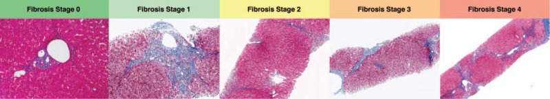
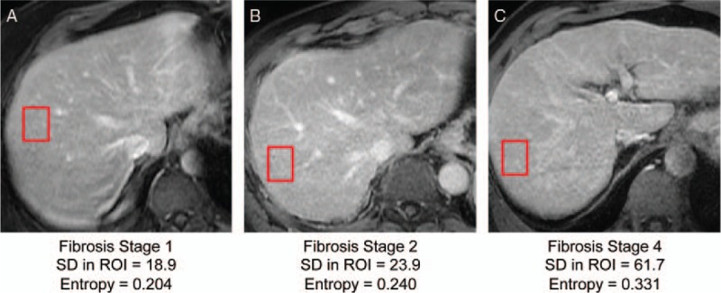
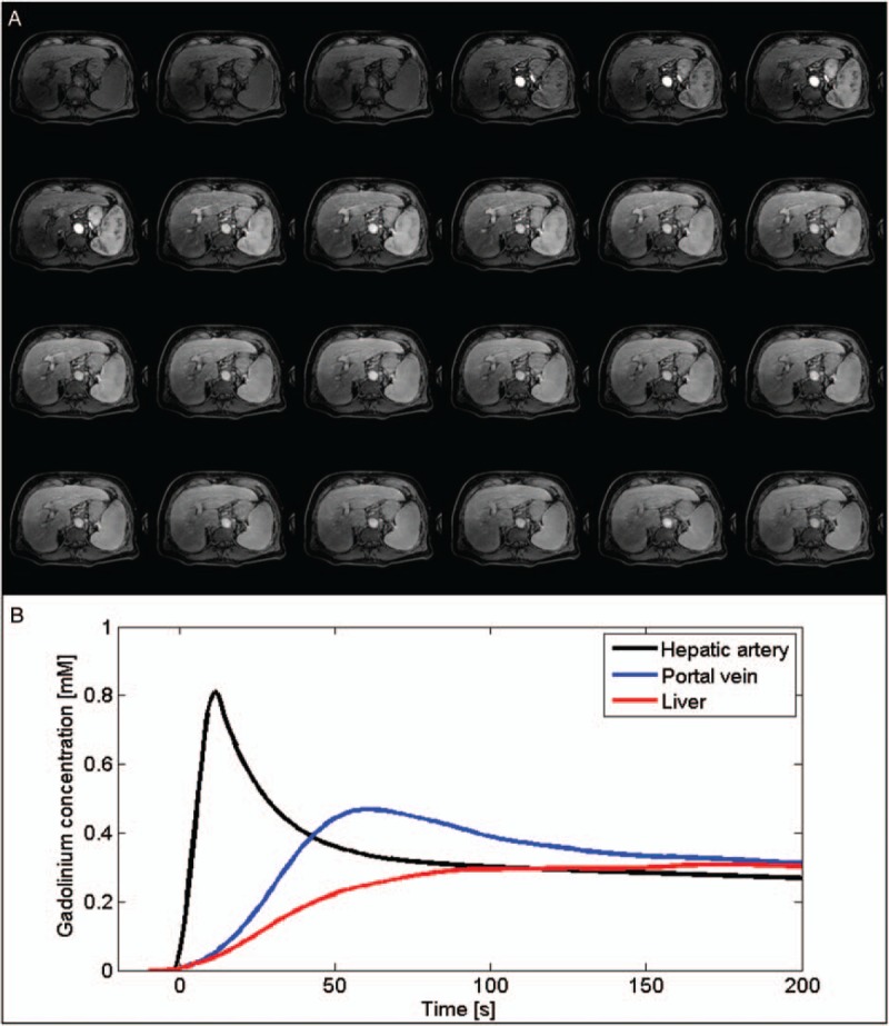
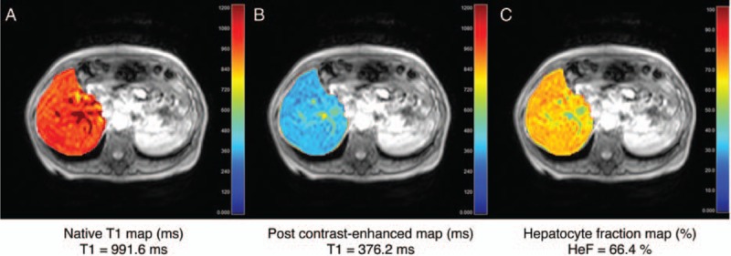
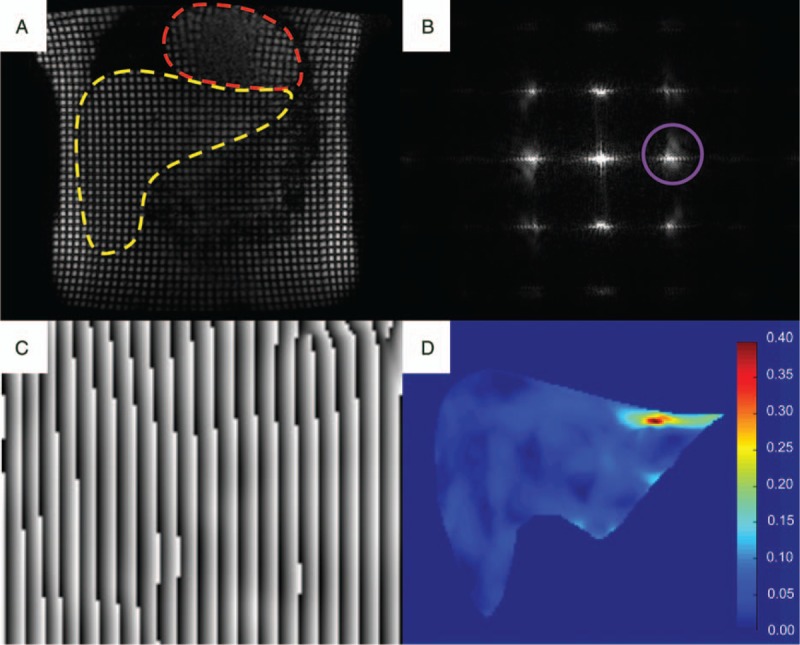
Liver fibrosis is a hallmark of chronic liver disease characterized by the excessive accumulation of extracellular matrix proteins. Although liver biopsy is the reference standard for diagnosis and staging of liver fibrosis, it has some limitations, including potential pain, sampling variability, and low patient acceptance. Hence, there has been an effort to develop noninvasive imaging techniques for diagnosis, staging, and monitoring of liver fibrosis. Many quantitative techniques have been implemented on magnetic resonance imaging (MRI) for this indication. The most widely validated technique is magnetic resonance elastography, which aims to measure viscoelastic properties of the liver and relate them to fibrosis stage. Several additional MRI methods have been developed or adapted to liver fibrosis quantification. Diffusion-weighted imaging measures the Brownian motion of water molecules which is restricted by collagen fibers. Texture analysis assesses the changes in the texture of liver parenchyma associated with fibrosis. Perfusion imaging relies on signal intensity and pharmacokinetic models to extract quantitative perfusion parameters. Hepatocellular function, which decreases with increasing fibrosis stage, can be estimated by the uptake of hepatobiliary contrast agents. Strain imaging measures liver deformation in response to physiological motion such as cardiac contraction. T1ρ quantification is an investigational technique, which measures the spin-lattice relaxation time in the rotating frame. This article will review the MRI techniques used in liver fibrosis staging, their advantages and limitations, and diagnostic performance. We will briefly discuss future directions, such as longitudinal monitoring of disease, prediction of portal hypertension, and risk stratification of hepatocellular carcinoma.
肝纤维化是慢性肝病的一个标志,其特征是细胞外基质蛋白过度积累。尽管肝活检是肝纤维化诊断和分期的参考标准,但它有一些局限性,包括潜在的疼痛、取样变异性和患者接受度低。因此,人们一直在努力开发用于肝纤维化诊断、分期和监测的非侵入性成像技术。许多定量技术已应用于磁共振成像(MRI)以用于此目的。最广泛验证的技术是磁共振弹性成像,其旨在测量肝脏的粘弹性特性并将其与纤维化阶段相关联。已经开发或改编了几种其他MRI方法用于肝纤维化定量。扩散加权成像测量受胶原纤维限制的水分子的布朗运动。纹理分析评估与纤维化相关的肝实质纹理变化。灌注成像依靠信号强度和药代动力学模型来提取定量灌注参数。随着纤维化阶段增加而降低的肝细胞功能可通过肝胆对比剂的摄取来估计。应变成像测量肝脏对诸如心脏收缩等生理运动的变形。T1ρ定量是一种研究技术,它测量旋转坐标系中的自旋晶格弛豫时间。本文将综述用于肝纤维化分期的MRI技术、它们的优点和局限性以及诊断性能。我们将简要讨论未来的方向,例如疾病的纵向监测、门静脉高压的预测以及肝细胞癌的风险分层。