• 文献检索
  • 文档翻译
  • 深度研究
  • 学术资讯
  • Suppr Zotero 插件Zotero 插件
  • 邀请有礼
  • 套餐&价格
  • 历史记录
应用&插件
Suppr Zotero 插件Zotero 插件浏览器插件Mac 客户端Windows 客户端微信小程序
定价
高级版会员购买积分包购买API积分包
服务
文献检索文档翻译深度研究API 文档MCP 服务
关于我们
关于 Suppr公司介绍联系我们用户协议隐私条款
关注我们

Suppr 超能文献

核心技术专利:CN118964589B侵权必究
粤ICP备2023148730 号-1Suppr @ 2026

文献检索

告别复杂PubMed语法,用中文像聊天一样搜索,搜遍4000万医学文献。AI智能推荐,让科研检索更轻松。

立即免费搜索

文件翻译

保留排版,准确专业,支持PDF/Word/PPT等文件格式,支持 12+语言互译。

免费翻译文档

深度研究

AI帮你快速写综述,25分钟生成高质量综述,智能提取关键信息,辅助科研写作。

立即免费体验

费舍尔评分系统和乳腺影像报告和数据系统在识别乳腺恶性病变中的准确性。

Accuracy of the Fischer scoring system and the Breast Imaging Reporting and Data System in identification of malignant breast lesions.

作者信息

Al-Khawari Hanaa, Athyal Reji, Kovacs Agnes, Al-Saleh Mervat, Madda John Patrick

机构信息

Department of Radiology, Faculty of Medicine, Kuwait University, 13110 Safat, Kuwait.

出版信息

Ann Saudi Med. 2009 Jul-Aug;29(4):280-7. doi: 10.4103/0256-4947.55310.

DOI:10.4103/0256-4947.55310
PMID:19584584
原文链接:https://pmc.ncbi.nlm.nih.gov/articles/PMC2841455/
Abstract

BACKGROUND AND OBJECTIVES

Fischer developed a scoring system in 1999 that made identifying malignnant lesions much easier for inexperienced radiologists. Our study was performed to assess whether this scoring system would help beginners to accurately diagnose breast lesions on magnetic resonance (MR) imaging and to assess the correlation between the magnetic resonance mammography Breast Imaging Reporting and Data System (MRM BI-RADS) grade and the final diagnosis.

PATIENTS AND METHODS

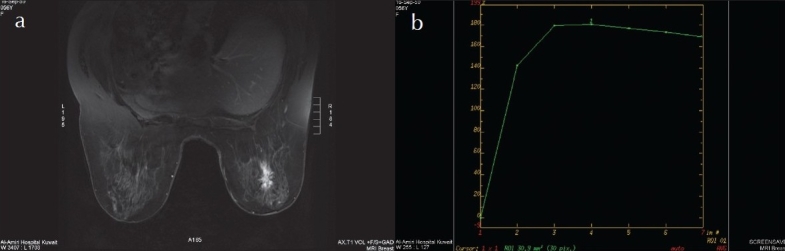
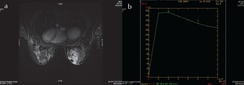
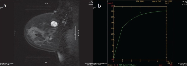
The lesion morphology and contrast kinetics of 63 masses in 41 patients were evaluated on MRI and accorded a MRM BI-RADS final assessment category using the Fischer scoring system. The accuracy was evaluated after the final diagnosis was obtained by tissue sampling and follow-up imaging.

RESULTS

There were 25 malignant and 30 benign lesions. Eight lesions were seen by MRI only and we could not verify their pathology since we did not have MR-guided biopsy facilities at the time of the study. On MR mammography, the proven carcinomatous lesions were characterized as BI-RADS category V in 16 (64%), category IV in 7 (28%), and category III in 2 (8%) lesions. Benign lesions were graded as category V in 3 (10%), category IV in 6 (20%), and category III in 3 (10%), category II in 10 (33%) and category I in 8 (27%) lesions. The MRM BI-RADS category accurately predicted malignancy in 92% and a benign pathology in 70% of the lesions. The overlap between the MRM features of chronic inflammatory lesions and carcinomas resulted in a lower accuracy in diagnosing benign as compared to malignant lesions.

CONCLUSION

The MRM BI-RADS lexicon using the Fischer scoring system is useful and has a high predictive value, especially for malignant breast lesions, and is easy to apply. Overlapping features between benign inflammatory and malignant lesions might yield a reduced accuracy in inflammatory pathologies.

摘要

背景与目的

1999年费舍尔开发了一种评分系统,这使得经验不足的放射科医生更容易识别恶性病变。我们开展这项研究是为了评估该评分系统是否有助于初学者在磁共振(MR)成像上准确诊断乳腺病变,并评估磁共振乳腺成像报告和数据系统(MRM BI-RADS)分级与最终诊断之间的相关性。

患者与方法

对41例患者的63个肿块的病变形态和对比剂动力学在MRI上进行评估,并使用费舍尔评分系统给予MRM BI-RADS最终评估类别。在通过组织取样和后续成像获得最终诊断后评估准确性。

结果

有25个恶性病变和30个良性病变。8个病变仅在MRI上可见,由于研究时我们没有MR引导活检设备,所以无法核实其病理情况。在MR乳腺成像上,经证实的癌性病变中,16个(64%)为BI-RADS V类,7个(28%)为IV类,2个(8%)为III类病变。良性病变中,3个(10%)为V类,6个(20%)为IV类,3个(10%)为III类,10个(33%)为II类,8个(27%)为I类病变。MRM BI-RADS类别在92%的病变中准确预测了恶性,在70%的病变中准确预测了良性病理。慢性炎性病变和癌的MRM特征之间的重叠导致诊断良性病变的准确性低于恶性病变。

结论

使用费舍尔评分系统的MRM BI-RADS词典是有用的,具有较高的预测价值,尤其是对于乳腺恶性病变,且易于应用。良性炎性病变和恶性病变之间的重叠特征可能导致炎性病变的诊断准确性降低。

https://cdn.ncbi.nlm.nih.gov/pmc/blobs/64c9/2841455/b535159a0cb2/ASM-29-280-g003.jpg
https://cdn.ncbi.nlm.nih.gov/pmc/blobs/64c9/2841455/abfcb0e12abb/ASM-29-280-g001.jpg
https://cdn.ncbi.nlm.nih.gov/pmc/blobs/64c9/2841455/d1a8d14c0697/ASM-29-280-g002.jpg
https://cdn.ncbi.nlm.nih.gov/pmc/blobs/64c9/2841455/b535159a0cb2/ASM-29-280-g003.jpg
https://cdn.ncbi.nlm.nih.gov/pmc/blobs/64c9/2841455/abfcb0e12abb/ASM-29-280-g001.jpg
https://cdn.ncbi.nlm.nih.gov/pmc/blobs/64c9/2841455/d1a8d14c0697/ASM-29-280-g002.jpg
https://cdn.ncbi.nlm.nih.gov/pmc/blobs/64c9/2841455/b535159a0cb2/ASM-29-280-g003.jpg

相似文献

1
Accuracy of the Fischer scoring system and the Breast Imaging Reporting and Data System in identification of malignant breast lesions.费舍尔评分系统和乳腺影像报告和数据系统在识别乳腺恶性病变中的准确性。
Ann Saudi Med. 2009 Jul-Aug;29(4):280-7. doi: 10.4103/0256-4947.55310.
2
Accuracy of the Fischer scoring system and the Breast Imaging Reporting and Data System in identification of malignant breast lesions.费舍尔评分系统与乳腺影像报告和数据系统在识别乳腺恶性病变中的准确性。
Hematol Oncol Stem Cell Ther. 2009;2(3):403-10. doi: 10.1016/s1658-3876(09)50009-x.
3
Positive predictive value of BI-RADS MR imaging.BI-RADS MR 成像的阳性预测值。
Radiology. 2012 Jul;264(1):51-8. doi: 10.1148/radiol.12110619. Epub 2012 May 15.
4
Dynamic contrast-enhanced MR imaging in screening detected microcalcification lesions of the breast: is there any value?动态对比增强磁共振成像在筛查乳腺微钙化病变中的应用:有价值吗?
Breast Cancer Res Treat. 2007 Jul;103(3):269-81. doi: 10.1007/s10549-006-9373-y. Epub 2006 Oct 25.
5
Contrast-enhanced MR mammography for evaluation of the contralateral breast in patients with diagnosed unilateral breast cancer or high-risk lesions.对比增强磁共振乳腺成像用于评估已诊断为单侧乳腺癌或高危病变患者的对侧乳房。
Radiology. 2007 Jun;243(3):670-80. doi: 10.1148/radiol.2433060838. Epub 2007 Apr 19.
6
MR imaging in probably benign lesions (BI-RADS category 3) of the breast.乳腺可能为良性病变(BI-RADS 3类)的磁共振成像
Eur J Radiol. 2006 Mar;57(3):436-44. doi: 10.1016/j.ejrad.2005.10.004. Epub 2005 Nov 28.
7
A comparative study of the diagnostic value of contrast-enhanced breast MR imaging and mammography on patients with BI-RADS 3-5 microcalcifications.对比增强乳腺磁共振成像与乳腺钼靶对BI-RADS 3-5级微钙化患者诊断价值的比较研究
PLoS One. 2014 Nov 3;9(11):e111217. doi: 10.1371/journal.pone.0111217. eCollection 2014.
8
A simplified scoring protocol to improve diagnostic accuracy with the breast imaging reporting and data system in breast magnetic resonance imaging.一种简化的评分方案,用于提高乳腺磁共振成像中乳腺影像报告和数据系统的诊断准确性。
Quant Imaging Med Surg. 2022 Jul;12(7):3860-3872. doi: 10.21037/qims-21-1036.
9
Cancer Yield and Patterns of Follow-up for BI-RADS Category 3 after Screening Mammography Recall in the National Mammography Database.在国家乳腺数据库中,乳腺 X 线摄影筛查召回后 BI-RADS 3 类的癌症发生率和随访模式。
Radiology. 2020 Jul;296(1):32-41. doi: 10.1148/radiol.2020192641. Epub 2020 May 19.
10
Role of magnetic resonance imaging in probably benign (BI-RADS category 3) microcalcifications of the breast.磁共振成像在乳腺可能良性(BI-RADS 类别 3)微钙化中的作用。
Radiol Med. 2014 Jun;119(6):393-9. doi: 10.1007/s11547-013-0361-0. Epub 2013 Dec 3.

引用本文的文献

1
Does contrast-enhanced ultrasound (CEUS) play a better role in diagnosis of breast lesions with calcification? A comparison with MRI.超声造影(CEUS)在钙化性乳腺病变的诊断中是否发挥着更好的作用?与磁共振成像(MRI)的比较。
Br J Radiol. 2020 Aug;93(1112):20200195. doi: 10.1259/bjr.20200195. Epub 2020 Jun 2.
2
Value of breast MRI for patients with a biopsy showing atypical ductal hyperplasia (ADH).乳腺 MRI 对活检显示非典型导管增生(ADH)患者的价值。
J Magn Reson Imaging. 2017 Dec;46(6):1738-1747. doi: 10.1002/jmri.25694. Epub 2017 Mar 10.

本文引用的文献

1
Breast magnetic resonance imaging: initial experience in Kuwait.
Med Princ Pract. 2009;18(2):143-8. doi: 10.1159/000189813. Epub 2009 Feb 10.
2
The current status of breast MR imaging. Part I. Choice of technique, image interpretation, diagnostic accuracy, and transfer to clinical practice.乳腺磁共振成像的现状。第一部分。技术选择、图像解读、诊断准确性及向临床实践的转化。
Radiology. 2007 Aug;244(2):356-78. doi: 10.1148/radiol.2442051620.
3
Breast imaging and reporting data system (BIRADS): magnetic resonance imaging.乳腺影像报告和数据系统(BIRADS):磁共振成像
Eur J Radiol. 2007 Feb;61(2):212-5. doi: 10.1016/j.ejrad.2006.08.036. Epub 2006 Dec 4.
4
Diagnostic architectural and dynamic features at breast MR imaging: multicenter study.乳腺磁共振成像的诊断性结构和动态特征:多中心研究
Radiology. 2006 Jan;238(1):42-53. doi: 10.1148/radiol.2381042117.
5
Screening with magnetic resonance imaging and mammography of a UK population at high familial risk of breast cancer: a prospective multicentre cohort study (MARIBS).对英国乳腺癌家族风险高的人群进行磁共振成像和乳房X线摄影筛查:一项前瞻性多中心队列研究(MARIBS)
Lancet. 2005;365(9473):1769-78. doi: 10.1016/S0140-6736(05)66481-1.
6
Problem solving MR imaging of the breast.乳腺问题的磁共振成像解决方法
Radiol Clin North Am. 2004 Sep;42(5):919-34, vii. doi: 10.1016/j.rcl.2004.05.001.
7
Breast carcinoma: effect of preoperative contrast-enhanced MR imaging on the therapeutic approach.乳腺癌:术前对比增强磁共振成像对治疗方法的影响。
Radiology. 1999 Dec;213(3):881-8. doi: 10.1148/radiology.213.3.r99dc01881.
8
Dynamic breast MR imaging: are signal intensity time course data useful for differential diagnosis of enhancing lesions?动态乳腺磁共振成像:信号强度时间过程数据对强化病变的鉴别诊断有用吗?
Radiology. 1999 Apr;211(1):101-10. doi: 10.1148/radiology.211.1.r99ap38101.
9
Improved diagnostic accuracy in dynamic contrast enhanced MRI of the breast by combined quantitative and qualitative analysis.
Br J Radiol. 1998 May;71(845):501-9. doi: 10.1259/bjr.71.845.9691895.
10
Diagnostic performance characteristics of architectural features revealed by high spatial-resolution MR imaging of the breast.乳腺高空间分辨率磁共振成像显示的结构特征的诊断性能特点
AJR Am J Roentgenol. 1997 Aug;169(2):409-15. doi: 10.2214/ajr.169.2.9242744.