Mohr P, Eggermont A M M, Hauschild A, Buzaid A
Elbekliniken, Buxtehude, Germany.
Ann Oncol. 2009 Aug;20 Suppl 6(Suppl 6):vi14-21. doi: 10.1093/annonc/mdp256.
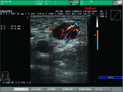
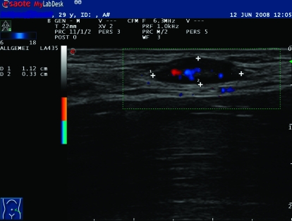
The American Joint Committee on Cancer (AJCC) staging of cutaneous melanoma is a continuously evolving system. The identification of increasingly more accurate prognostic factors has led to major changes in melanoma staging over the years, and the current system described in this review will likely be modified in the near future. Likewise, application of new imaging techniques has also changed the staging work-up of patients with cutaneous melanoma. Chest and abdominal computed tomography (CT) scanning is most commonly used for evaluation of potential metastatic sites in the lungs, lymph nodes and liver, and is indicated in patients with new symptoms, anaemia, elevated lactate dehydrogenase or a chest X-ray abnormality. CT scans should be restricted to patients with high-risk melanoma (stage IIC, IIIB, IIIC and stage IIIA with a macroscopic sentinel lymph node). Magnetic resonance imaging (MRI) of the brain is a mandatory test in patients with stage IV, optional in stage III and not used in patients with stage I and II disease. Positron emission tomography (PET)/CT is more accurate than CT or MRI alone in the diagnosis of metastases and should complement conventional CT/MRI imaging in the staging work-up of patients who have solitary or oligometastatic disease where surgical resection is most relevant.
美国癌症联合委员会(AJCC)的皮肤黑色素瘤分期是一个不断发展的系统。多年来,越来越准确的预后因素的确定导致了黑色素瘤分期的重大变化,本综述中描述的当前系统在不久的将来可能会被修改。同样,新成像技术的应用也改变了皮肤黑色素瘤患者的分期检查。胸部和腹部计算机断层扫描(CT)最常用于评估肺部、淋巴结和肝脏的潜在转移部位,适用于有新症状、贫血、乳酸脱氢酶升高或胸部X线异常的患者。CT扫描应限于高危黑色素瘤患者(II期C、IIIB期、IIIC期以及有肉眼可见前哨淋巴结的IIIA期)。对于IV期患者,脑部磁共振成像(MRI)是一项必需的检查,III期患者可选择进行,而I期和II期患者则不使用。在转移灶的诊断方面,正电子发射断层扫描(PET)/CT比单纯的CT或MRI更准确,对于有孤立性或寡转移性疾病且手术切除最为关键的患者,在分期检查中应作为传统CT/MRI成像的补充。