Ohnishi Takayuki, Matsumura Shinji, Ito Seiji
Department of Medical Chemistry, Kansai Medical University, Fumizono, Moriguchi, Japan.
Mol Pain. 2009 Jul 20;5:40. doi: 10.1186/1744-8069-5-40.
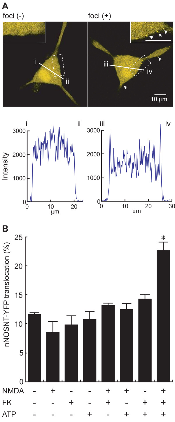
The translocation of neuronal nitric oxide synthase (nNOS) from the cytosol to the membrane is functionally coupled to the activation of N-methyl-D-aspartate (NMDA) receptors at synapses. Whereas there is abundant evidence indicating that ATP and nitric oxide are involved in nociceptive transmission, whether nNOS is activated by ATP remains unknown. We recently established a fluorescence imaging system for examining nNOS translocation in PC12 cells expressing a yellow fluorescence protein-tagged nNOS N-terminal mutant, nNOSNT-YFP, and examined the effect of ATP on nNOS translocation using the system.
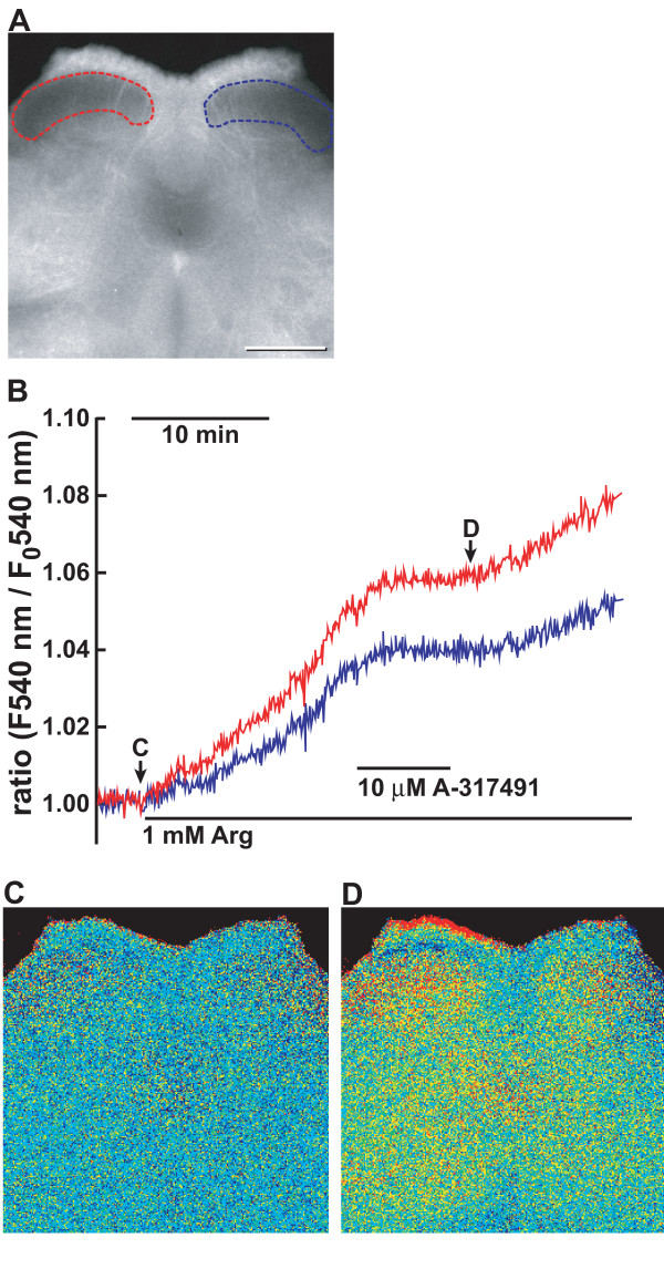
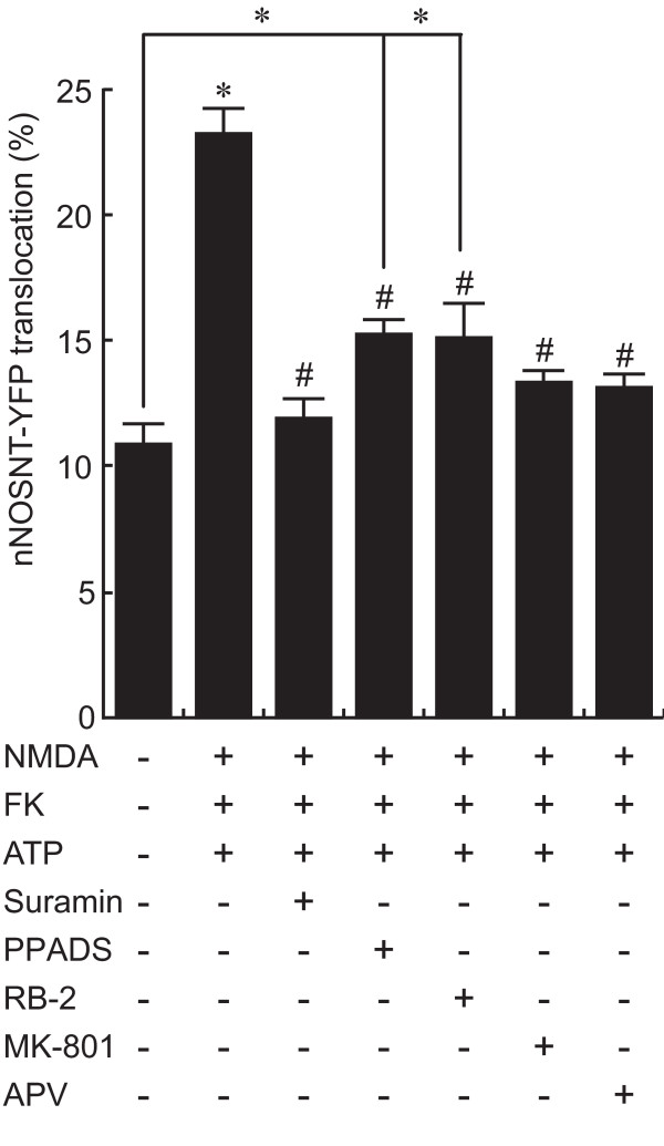
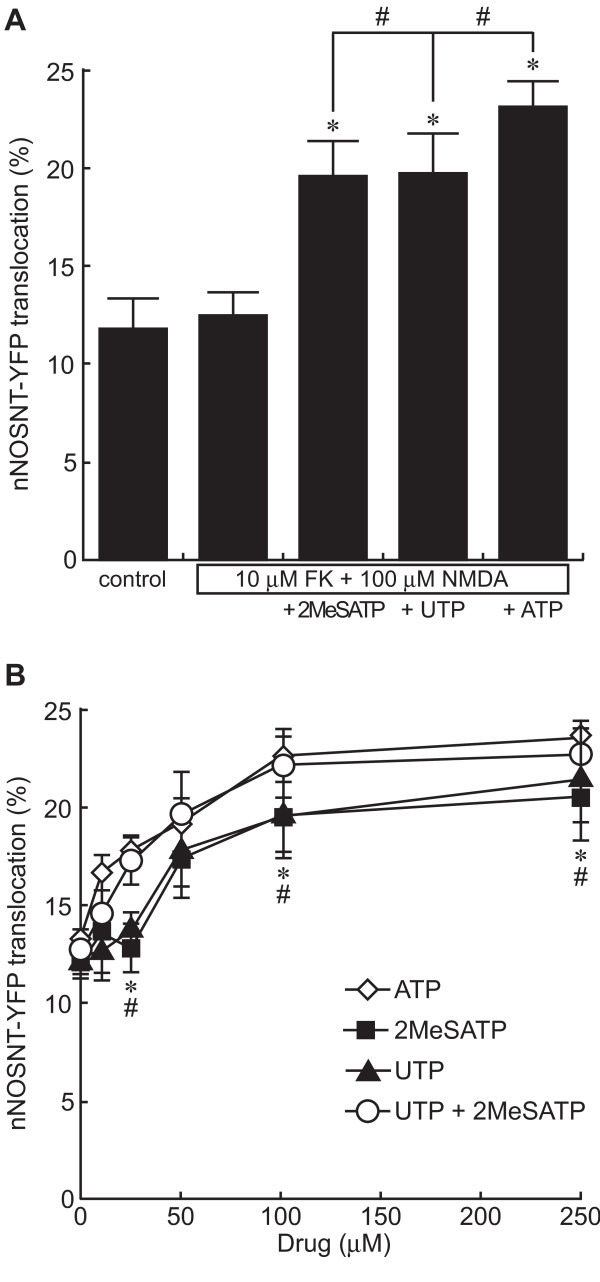
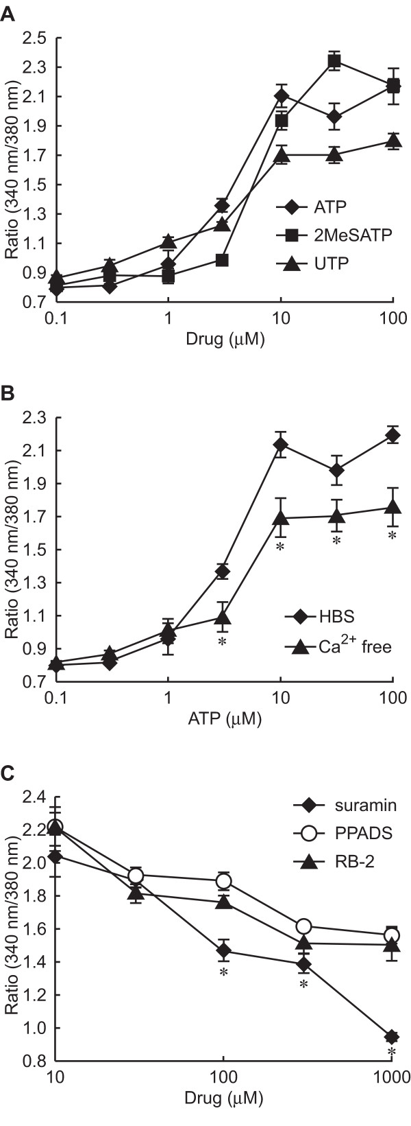
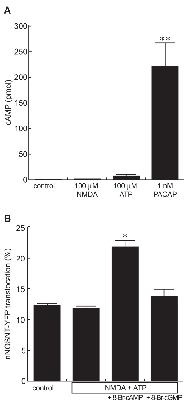
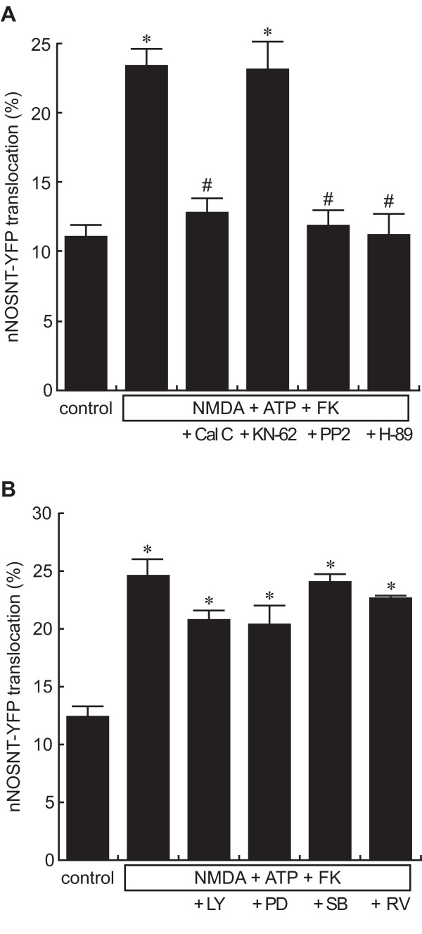
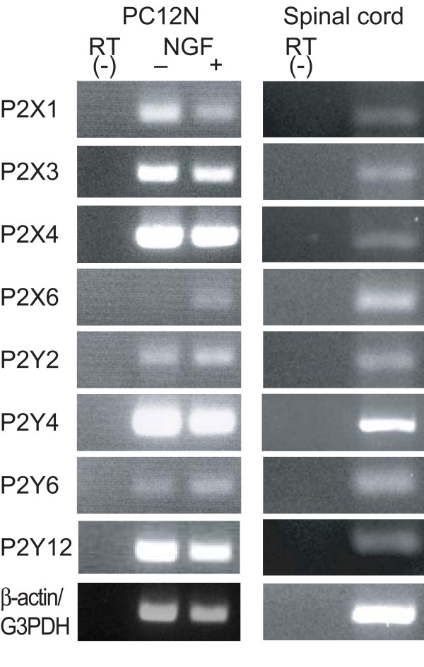
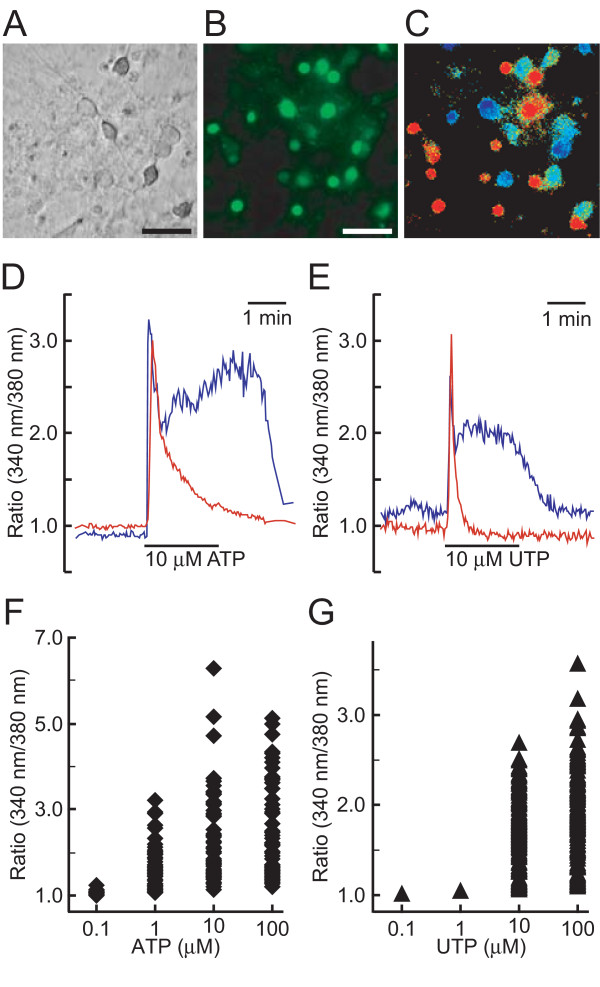
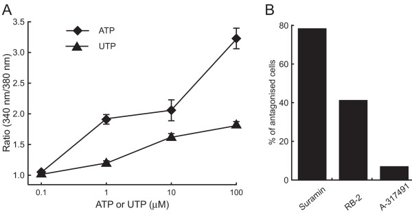
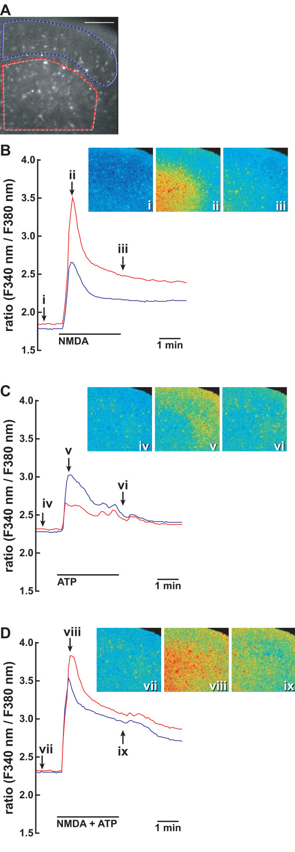
The translocation of nNOS was induced by ATP in the presence of NMDA and forskolin, an adenylate cyclase activator. The purinergic P2X receptor agonist 2-MeSATP and the P2Y agonist UTP significantly enhanced nNOS translocation; and simultaneous stimulation with 2-MeSATP and UTP exhibited the same concentration-response curve for the translocation as obtained with ATP. ATP, 2-MeSATP, and UTP increased the intracellular Ca2+ concentration ([Ca2+]i) in PC12 cells. Conversely, whereas the P2X receptor antagonist PPADS and the P2Y antagonist reactive blue-2 partially inhibited increases in the translocation of nNOS and [Ca2+]i by ATP, the non-selective P2 receptor antagonist suramin completely blocked them. In addition, the increase in the nNOS translocation by ATP was blocked by NMDA receptor antagonists and inhibitors of protein kinase A, protein kinase C, and Src kinase. Consistent with the expression of P2X and P2Y receptors in the spinal cord, ATP and UTP increased the [Ca2+]i in primary cultured spinal neurons. ATP potentiated and prolonged the [Ca2+]i increase produced by NMDA in the dorsal horn of the spinal cord. Furthermore, the selective P2X3/P2X2/3 antagonist A-317491 inhibited nNOS activation assessed by NO formation in spinal slices prepared from neuropathic pain model mice.
ATP is involved in nNOS translocation mediated by protein kinase C via activation of P2X and P2Y receptors and nNOS translocation may be an action mechanism of ATP in nocieptive processing in the spinal cord.
神经元型一氧化氮合酶(nNOS)从胞质溶胶向细胞膜的转位在功能上与突触处N-甲基-D-天冬氨酸(NMDA)受体的激活相关联。尽管有大量证据表明ATP和一氧化氮参与伤害性感受传递,但nNOS是否被ATP激活仍不清楚。我们最近建立了一种荧光成像系统,用于检测表达黄色荧光蛋白标记的nNOS N端突变体(nNOSNT-YFP)的PC12细胞中nNOS的转位,并使用该系统检测了ATP对nNOS转位的影响。
在NMDA和腺苷酸环化酶激活剂福斯可林存在的情况下,ATP可诱导nNOS转位。嘌呤能P2X受体激动剂2-甲基硫代ATP(2-MeSATP)和P2Y激动剂UTP显著增强nNOS转位;同时用2-MeSATP和UTP刺激产生的转位浓度-反应曲线与ATP相同。ATP、2-MeSATP和UTP可增加PC12细胞内的Ca2+浓度([Ca2+]i)。相反,P2X受体拮抗剂吡哆醛-6-偶氮(苯-2,4-二磺酸)二钠盐(PPADS)和P2Y拮抗剂反应性蓝-2可部分抑制ATP诱导的nNOS转位和[Ca2+]i增加,而非选择性P2受体拮抗剂苏拉明则完全阻断它们。此外,ATP诱导的nNOS转位增加被NMDA受体拮抗剂以及蛋白激酶A、蛋白激酶C和Src激酶的抑制剂所阻断。与脊髓中P2X和P2Y受体的表达一致,ATP和UTP可增加原代培养脊髓神经元中的[Ca2+]i。ATP增强并延长了NMDA在脊髓背角产生的[Ca2+]i增加。此外,选择性P2X3/P2X2/3拮抗剂A-317491可抑制通过对神经性疼痛模型小鼠制备的脊髓切片中一氧化氮生成评估的nNOS激活。
ATP通过激活P2X和P2Y受体参与蛋白激酶C介导的nNOS转位,nNOS转位可能是ATP在脊髓伤害性感受处理中的一种作用机制。