Zalaudek Iris, Docimo Giovanni, Argenziano Giuseppe
Medical University of Graz, Austria.
Arch Dermatol. 2009 Jul;145(7):816-26. doi: 10.1001/archdermatol.2009.115.
To review recent dermoscopy studies that provide new insights into the evolution of nevi and their patterns of pigmentation as they contribute to the diagnosis of nevi and the management of pigmented melanocytic nevi.
Data for this article were identified by searching the English and German literature by Medline and Journals@Ovid search for the period 1950 to January 2009.
The following relevant terms were used: dermoscopy, dermatoscopy, epiluminescence microscopy (ELM), surface microscopy, digital dermoscopy, digital dermatoscopy, digital epiluminescence microscopy, digital surface microscopy, melanocytic skin lesion, nevi, and pigmented skin lesions. There were no exclusion criteria.
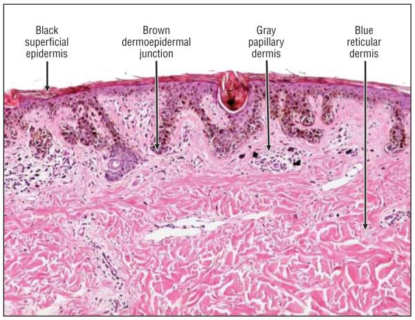
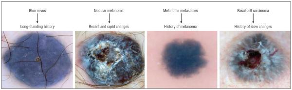
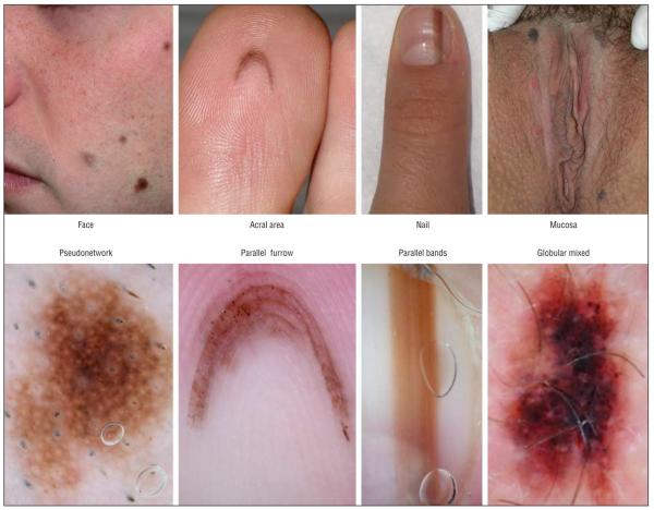
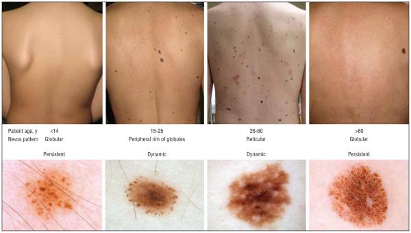
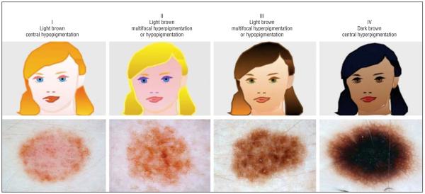
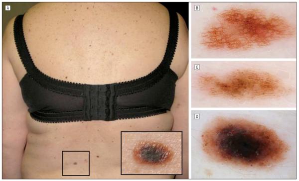
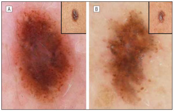
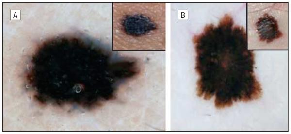
The dermoscopic diagnosis of nevi relies on the following 4 criteria (each of which is characterized by 4 variables): (1) color (black, brown, gray, and blue); (2) pattern (globular, reticular, starburst, and homogeneous blue pattern); (3) pigment distribution (multifocal, central, eccentric, and uniform); and (4) special sites (face, acral areas, nail, and mucosa). In addition, the following 6 factors related to the patient might influence the pattern of pigmentation of the individual nevi: age, skin type, history of melanoma, UV exposure, pregnancy, and growth dynamics.
The 4 x 4 x 6 "rule" may help clinicians remember the basic dermoscopic criteria of nevi and the patient-related factors influencing their patterns. Dermoscopy is a useful technique for diagnosing melanocytic nevi, but the clinician should take additional factors into consideration to optimize the management of cases of pigmented lesions.
回顾近期的皮肤镜研究,这些研究为痣的演变及其色素沉着模式提供了新的见解,有助于痣的诊断及色素性黑素细胞痣的管理。
通过检索1950年至2009年1月期间的英文和德文文献,利用医学文献数据库(Medline)和Ovid期刊数据库确定本文的数据。
使用了以下相关术语:皮肤镜检查、真皮镜检查、表皮透光显微镜检查(ELM)、表面显微镜检查、数字皮肤镜检查、数字真皮镜检查、数字表皮透光显微镜检查、数字表面显微镜检查、黑素细胞性皮肤病变、痣和色素性皮肤病变。没有排除标准。
痣的皮肤镜诊断依赖于以下4条标准(每条标准由4个变量表征):(1)颜色(黑色、棕色、灰色和蓝色);(2)模式(球状、网状、星芒状和均匀蓝色模式);(3)色素分布(多灶性、中央性、偏心性和均匀性);(4)特殊部位(面部、肢端、指甲和黏膜)。此外,以下与患者相关的6个因素可能影响单个痣的色素沉着模式:年龄、皮肤类型、黑色素瘤病史、紫外线暴露、妊娠和生长动态。
4×4×6“规则”可能有助于临床医生记住痣的基本皮肤镜标准以及影响其模式的患者相关因素。皮肤镜检查是诊断黑素细胞痣的一项有用技术,但临床医生应考虑其他因素以优化色素性病变病例的管理。