• 文献检索
  • 文档翻译
  • 深度研究
  • 学术资讯
  • Suppr Zotero 插件Zotero 插件
  • 邀请有礼
  • 套餐&价格
  • 历史记录
应用&插件
Suppr Zotero 插件Zotero 插件浏览器插件Mac 客户端Windows 客户端微信小程序
定价
高级版会员购买积分包购买API积分包
服务
文献检索文档翻译深度研究API 文档MCP 服务
关于我们
关于 Suppr公司介绍联系我们用户协议隐私条款
关注我们

Suppr 超能文献

核心技术专利:CN118964589B侵权必究
粤ICP备2023148730 号-1Suppr @ 2026

文献检索

告别复杂PubMed语法,用中文像聊天一样搜索,搜遍4000万医学文献。AI智能推荐,让科研检索更轻松。

立即免费搜索

文件翻译

保留排版,准确专业,支持PDF/Word/PPT等文件格式,支持 12+语言互译。

免费翻译文档

深度研究

AI帮你快速写综述,25分钟生成高质量综述,智能提取关键信息,辅助科研写作。

立即免费体验

原发性颅外脑膜瘤:146例分析

Primary extracranial meningiomas: an analysis of 146 cases.

作者信息

Rushing Elisabeth J, Bouffard John-Paul, McCall Sherman, Olsen Cara, Mena Hernando, Sandberg Glenn D, Thompson Lester D R

机构信息

Department of Neuropathology and Ophthalmic Pathology, Armed Forces Institute of Pathology, Washington, DC 20306-6000, USA.

出版信息

Head Neck Pathol. 2009 Jun;3(2):116-30. doi: 10.1007/s12105-009-0118-1. Epub 2009 May 20.

DOI:10.1007/s12105-009-0118-1
PMID:19644540
原文链接:https://pmc.ncbi.nlm.nih.gov/articles/PMC2715454/
Abstract

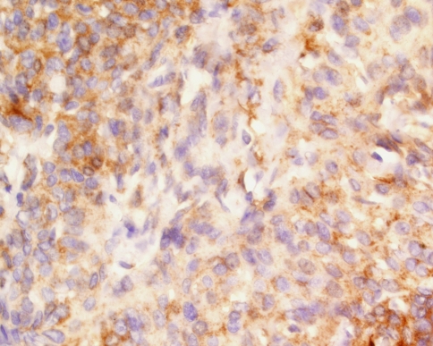
Primary extracranial meningiomas are rare neoplasms, frequently misdiagnosed, resulting in inappropriate clinical management. To date, a large clinicopathologic study has not been reported. One hundred and forty-six cases diagnosed between 1970 and 1999 were retrieved from the files of the Armed Forces Institute of Pathology. Histologic features were reviewed, immunohistochemistry analysis was performed (n = 85), and patient follow-up was obtained (n = 110). The patients included 74 (50.7%) females and 72 (49.3%) males. Tumors of the skin were much more common in males than females (1.7:1). There was an overall mean age at presentation of 42.4 years, with a range of 0.3-88 years. The overall mean age at presentation was significantly younger for skin primaries (36.2 years) than for ear (50.1 years) and nasal cavity (47.1 years) primaries. Symptoms were in general non-specific and reflected the anatomic site of involvement, affecting the following areas in order of frequency: scalp skin (40.4%), ear and temporal bone (26%), and sinonasal tract (24%). The tumors ranged in size from 0.5 up to 8 cm, with a mean size of 2.3 cm. Histologically, the majority of tumors were meningothelial (77.4%), followed by atypical (7.5%), psammomatous (4.1%) and anaplastic (2.7%). Psammoma bodies were present in 45 tumors (30.8%), and bone invasion in 31 (21.2%) of tumors. The vast majority were WHO Grade I tumors (87.7%), followed by Grade II (9.6%) and Grade III (2.7%) tumors. Immunohistochemically, the tumor cells labeled for EMA (76%; 61/80), S-100 protein (19%; 15/78), CK 7 (22%; 12/55), and while there was ki-67 labeling in 27% (21/78), <3% of cells were positive. The differential diagnosis included a number of mesenchymal and epithelial tumors (paraganglioma, schwannoma, carcinoma, melanoma, neuroendocrine adenoma of the middle ear), depending on the anatomic site of involvement. Treatment and follow-up was available in 110 patients: Biopsy, local excision, or wide excision was employed. Follow-up time ranged from 1 month to 32 years, with an average of 14.5 years. Recurrences were noted in 26 (23.6%) patients, who were further managed by additional surgery. At last follow-up, recurrent disease was persistent in 15 patients (mean, 7.7 years): 13 patients were dead (died with disease) and two were alive; the remaining patients were disease free (alive 60, mean 19.0 years, dead 35, mean 9.6 years). There is no statistically significant difference in 5-year survival rates by site: ear and temporal bone: 83.3%; nasal cavity: 81.8%; scalp skin: 78.5%; other sites: 65.5% (P = 0.155). Meningiomas can present in a wide variety of sites, especially within the head and neck region. They behave as slow-growing neoplasms with a good prognosis, with longest survival associated with younger age, and complete resection. Awareness of this diagnosis in an unexpected location will help to avoid potential difficulties associated with the diagnosis and management of these tumors.

https://cdn.ncbi.nlm.nih.gov/pmc/blobs/78f1/2811612/cf54ab140361/12105_2009_118_Fig2_HTML.jpg
https://cdn.ncbi.nlm.nih.gov/pmc/blobs/78f1/2811612/08d80d205941/12105_2009_118_Fig1_HTML.jpg
https://cdn.ncbi.nlm.nih.gov/pmc/blobs/78f1/2811612/cf54ab140361/12105_2009_118_Fig2_HTML.jpg
https://cdn.ncbi.nlm.nih.gov/pmc/blobs/78f1/2811612/08d80d205941/12105_2009_118_Fig1_HTML.jpg
https://cdn.ncbi.nlm.nih.gov/pmc/blobs/78f1/2811612/cf54ab140361/12105_2009_118_Fig2_HTML.jpg
摘要

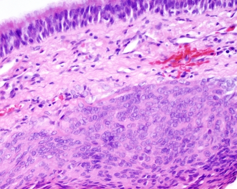
原发性颅外脑膜瘤是罕见的肿瘤,常被误诊,导致临床处理不当。迄今为止,尚未见大型临床病理研究报道。从武装部队病理研究所档案中检索出1970年至1999年间诊断的146例病例。回顾组织学特征,进行免疫组化分析(n = 85),并获取患者随访资料(n = 110)。患者包括74名女性(50.7%)和72名男性(49.3%)。男性皮肤肿瘤比女性更常见(1.7:1)。总体就诊时平均年龄为42.4岁,范围为0.3 - 88岁。皮肤原发性肿瘤的总体就诊平均年龄(36.2岁)明显低于耳部(50.1岁)和鼻腔(47.1岁)原发性肿瘤。症状一般无特异性,反映受累的解剖部位,按频率依次影响以下部位:头皮皮肤(40.4%)、耳和颞骨(26%)、鼻窦道(24%)。肿瘤大小从0.5厘米至8厘米不等,平均大小为2.3厘米。组织学上,大多数肿瘤为脑膜皮型(77.4%),其次是非典型型(7.5%)、砂粒体型(4.1%)和间变型(2.7%)。45个肿瘤(30.8%)有砂粒体,31个肿瘤(21.2%)有骨质侵犯。绝大多数为世界卫生组织I级肿瘤(87.7%),其次是II级(9.6%)和III级(2.7%)肿瘤。免疫组化方面,肿瘤细胞EMA标记阳性率为76%(61/80),S - 100蛋白为19%(15/78),CK 7为22%(12/55),ki - 67标记阳性率为27%(21/78),<3%的细胞呈阳性。鉴别诊断包括一些间叶性和上皮性肿瘤(副神经节瘤、神经鞘瘤、癌、黑色素瘤、中耳神经内分泌腺瘤),取决于受累的解剖部位。110例患者有治疗及随访资料:采用活检、局部切除或广泛切除。随访时间从1个月至32年不等,平均为14.5年。26例(23.6%)患者出现复发,进一步行手术治疗。末次随访时,15例患者复发疾病持续存在(平均7.7年):13例患者死亡(死于疾病),2例存活;其余患者无疾病(存活60例,平均19.0年,死亡35例,平均9.6年)。各部位5年生存率无统计学显著差异:耳和颞骨:83.3%;鼻腔:81.8%;头皮皮肤:78.5%;其他部位:65.5%(P = 0.155)。脑膜瘤可出现在多种部位,尤其是头颈部区域。它们表现为生长缓慢的肿瘤,预后良好,年龄越小、完全切除者生存期最长。在意外部位意识到这一诊断将有助于避免与这些肿瘤的诊断和处理相关的潜在困难。

相似文献

1
Primary extracranial meningiomas: an analysis of 146 cases.原发性颅外脑膜瘤:146例分析
Head Neck Pathol. 2009 Jun;3(2):116-30. doi: 10.1007/s12105-009-0118-1. Epub 2009 May 20.
2
Primary ear and temporal bone meningiomas: a clinicopathologic study of 36 cases with a review of the literature.原发性耳及颞骨脑膜瘤:36例临床病理研究并文献复习
Mod Pathol. 2003 Mar;16(3):236-45. doi: 10.1097/01.MP.0000056631.15739.1B.
3
Extracranial sinonasal tract meningiomas: a clinicopathologic study of 30 cases with a review of the literature.颅外鼻窦道脑膜瘤:30例临床病理研究并文献复习
Am J Surg Pathol. 2000 May;24(5):640-50. doi: 10.1097/00000478-200005000-00002.
4
Sinonasal tract mucoepidermoid carcinoma: a clinicopathologic and immunophenotypic study of 19 cases combined with a comprehensive review of the literature.鼻窦黏液表皮样癌:19例临床病理及免疫表型研究并文献综合复习
Head Neck Pathol. 2012 Jun;6(2):191-207. doi: 10.1007/s12105-011-0320-9. Epub 2011 Dec 20.
5
Sinonasal tract and nasopharyngeal adenoid cystic carcinoma: a clinicopathologic and immunophenotypic study of 86 cases.鼻窦和鼻咽腺样囊性癌:86例临床病理及免疫表型研究
Head Neck Pathol. 2014 Mar;8(1):88-109. doi: 10.1007/s12105-013-0487-3. Epub 2013 Sep 15.
6
Ectopic sphenoid sinus pituitary adenoma (ESSPA) with normal anterior pituitary gland: a clinicopathologic and immunophenotypic study of 32 cases with a comprehensive review of the english literature.垂体前叶正常的异位蝶窦垂体腺瘤(ESSPA):32例临床病理及免疫表型研究并对英文文献进行全面综述
Head Neck Pathol. 2012 Mar;6(1):75-100. doi: 10.1007/s12105-012-0336-9. Epub 2012 Mar 20.
7
Sinonasal tract angiosarcoma: a clinicopathologic and immunophenotypic study of 10 cases with a review of the literature.鼻窦血管肉瘤:10例临床病理及免疫表型研究并文献复习
Head Neck Pathol. 2007 Sep;1(1):1-12. doi: 10.1007/s12105-007-0017-2. Epub 2007 Oct 25.
8
Primary extradural meningiomas in head: a report of 19 cases and review of literature.原发性颅内硬脑膜外脑膜瘤:19例报告并文献复习
Int J Clin Exp Pathol. 2015 May 1;8(5):5624-32. eCollection 2015.
9
Ki-67 proliferative index predicts clinical outcome in patients with atypical or anaplastic meningioma.Ki-67增殖指数可预测非典型或间变性脑膜瘤患者的临床预后。
Neuropathology. 2007 Apr;27(2):114-20. doi: 10.1111/j.1440-1789.2007.00750.x.
10
Perivascular epithelioid cell neoplasms of soft tissue and gynecologic origin: a clinicopathologic study of 26 cases and review of the literature.软组织和妇科来源的血管周上皮样细胞肿瘤:26例临床病理研究及文献复习
Am J Surg Pathol. 2005 Dec;29(12):1558-75. doi: 10.1097/01.pas.0000173232.22117.37.

引用本文的文献

1
Unusual adrenal metastasis of anaplastic meningioma: A case report.间变性脑膜瘤罕见的肾上腺转移:一例报告
Int J Surg Case Rep. 2025 Jul 20;134:111707. doi: 10.1016/j.ijscr.2025.111707.
2
An Extracranial, Infratemporal Meningioma Following Neoadjuvant Radiotherapy: A Case Report.新辅助放疗后颅外颞下窝脑膜瘤:一例报告
Cureus. 2025 Jun 23;17(6):e86612. doi: 10.7759/cureus.86612. eCollection 2025 Jun.
3
A Rare Case of Primary Pulmonary Meningioma.原发性肺脑膜瘤1例罕见病例

本文引用的文献

1
Extracranial meningioma in the vicinity of the temporal bone: a difficult preoperative diagnosis.颞骨附近的颅外脑膜瘤:术前诊断困难
Surg Neurol. 2007 Jan;67(1):102-5. doi: 10.1016/j.surneu.2006.02.043. Epub 2006 Oct 4.
2
Extracranial meningioma presenting as a neck mass in a patient with underlying ankylosing spondylitis.在一名患有强直性脊柱炎的患者中,颅外脑膜瘤表现为颈部肿块。
J Rheumatol. 2006 Sep;33(9):1883-5.
3
Primary extracranial meningioma of the pelvis: a light microscopic, immunohistochemical, and ultrastructural study.
J Clin Med. 2025 Apr 15;14(8):2688. doi: 10.3390/jcm14082688.
4
Cutaneous meningothelial proliferative lesions of the parietal skin in eleven cats.11只猫顶叶皮肤的皮肤脑膜上皮增生性病变
J Vet Med Sci. 2025 May 15;87(5):546-550. doi: 10.1292/jvms.24-0386. Epub 2025 Mar 28.
5
Primary Extracranial Meningiomas of the Sinonasal Tract: A Systematic Review.鼻窦原发性颅外脑膜瘤:一项系统评价
J Neurol Surg B Skull Base. 2024 Mar 8;86(1):112-121. doi: 10.1055/a-2263-1881. eCollection 2025 Feb.
6
Palato-Antral Involvement of a Primary Extracranial Sinonasal Meningioma.原发性颅外鼻窦脑膜瘤的腭窦受累情况。
Sultan Qaboos Univ Med J. 2024 Nov;24(4):575-577. doi: 10.18295/squmj.12.2023.096. Epub 2024 Nov 27.
7
Benign extracranial meningioma with pulmonary metastasis: a case report and review of literature.良性颅外脑膜瘤伴肺转移:病例报告及文献复习。
J Med Case Rep. 2024 Oct 5;18(1):461. doi: 10.1186/s13256-024-04800-z.
8
Primary Typical Meningioma of the Sphenoid Sinus: A Case Report and Literature Review.蝶窦原发性典型脑膜瘤:一例报告及文献复习
Indian J Otolaryngol Head Neck Surg. 2024 Aug;76(4):3722-3725. doi: 10.1007/s12070-024-04722-8. Epub 2024 Apr 26.
9
Primary pulmonary meningioma and minute pulmonary meningothelial-like nodules: Rare pulmonary nodular lesions requiring more awareness in clinical practice.原发性肺脑膜瘤和微小肺脑膜上皮样结节:临床实践中需要更多关注的罕见肺结节性病变。
World J Clin Cases. 2024 Apr 16;12(11):1857-1862. doi: 10.12998/wjcc.v12.i11.1857.
10
FNA of Meningioma with Rhabdoid Features Presenting as a Lateral Neck Mass.脑膜瘤伴横纹肌样特征的细针吸取细胞学检查,表现为侧颈部肿块。
Head Neck Pathol. 2024 Apr 24;18(1):32. doi: 10.1007/s12105-024-01644-3.
Gynecol Oncol. 2006 Nov;103(2):745-8. doi: 10.1016/j.ygyno.2006.05.050. Epub 2006 Aug 9.
4
Intraventricular meninigiomas: a report of 25 cases.脑室内脑膜瘤:25例报告。
Neurosurg Rev. 2006 Jan;29(1):36-40. doi: 10.1007/s10143-005-0418-1. Epub 2005 Oct 12.
5
Primary extracranial meningioma of the sinonasal tract.鼻窦原发性颅外脑膜瘤。
Acta Radiol. 2005 Jul;46(4):415-8. doi: 10.1080/02841850510021210.
6
Expression of cytokeratin by malignant meningiomas: diagnostic pitfall of cytokeratin to separate malignant meningiomas from metastatic carcinoma.细胞角蛋白在恶性脑膜瘤中的表达:细胞角蛋白用于区分恶性脑膜瘤与转移性癌的诊断陷阱。
Mod Pathol. 2004 Sep;17(9):1129-33. doi: 10.1038/modpathol.3800162.
7
Primary extracranial meningioma in the vicinity of the temporal bone: a benign lesion which is rarely recognized clinically.颞骨附近的原发性颅外脑膜瘤:一种临床上很少被识别的良性病变。
Acta Otolaryngol. 2004 Jan;124(1):5-7. doi: 10.1080/00016480310014903.
8
Extracranial meningioma presenting as a tumor of the neck.表现为颈部肿瘤的颅外脑膜瘤。
Am J Surg. 1960 Sep;100:486-9. doi: 10.1016/0002-9610(60)90394-9.
9
Cutaneous meningioma (psammoma); report of a case.
AMA Arch Derm. 1956 Dec;74(6):590-4. doi: 10.1001/archderm.1956.01550120010002.
10
Sinonasal tract and nasopharyngeal melanomas: a clinicopathologic study of 115 cases with a proposed staging system.鼻窦和鼻咽黑色素瘤:115例临床病理研究及拟用分期系统
Am J Surg Pathol. 2003 May;27(5):594-611. doi: 10.1097/00000478-200305000-00004.