• 文献检索
  • 文档翻译
  • 深度研究
  • 学术资讯
  • Suppr Zotero 插件Zotero 插件
  • 邀请有礼
  • 套餐&价格
  • 历史记录
应用&插件
Suppr Zotero 插件Zotero 插件浏览器插件Mac 客户端Windows 客户端微信小程序
定价
高级版会员购买积分包购买API积分包
服务
文献检索文档翻译深度研究API 文档MCP 服务
关于我们
关于 Suppr公司介绍联系我们用户协议隐私条款
关注我们

Suppr 超能文献

核心技术专利:CN118964589B侵权必究
粤ICP备2023148730 号-1Suppr @ 2026

文献检索

告别复杂PubMed语法,用中文像聊天一样搜索,搜遍4000万医学文献。AI智能推荐,让科研检索更轻松。

立即免费搜索

文件翻译

保留排版,准确专业,支持PDF/Word/PPT等文件格式,支持 12+语言互译。

免费翻译文档

深度研究

AI帮你快速写综述,25分钟生成高质量综述,智能提取关键信息,辅助科研写作。

立即免费体验

在使用部分体积图的拉格朗日 - 欧拉偏微分方程组合方法中基于体素的自动三维皮质厚度测量。

Automated voxel-based 3D cortical thickness measurement in a combined Lagrangian-Eulerian PDE approach using partial volume maps.

作者信息

Acosta Oscar, Bourgeat Pierrick, Zuluaga Maria A, Fripp Jurgen, Salvado Olivier, Ourselin Sébastien

机构信息

The Australian e-Health Research Centre, CSIRO ICT Centre, Brisbane, Australia.

出版信息

Med Image Anal. 2009 Oct;13(5):730-43. doi: 10.1016/j.media.2009.07.003. Epub 2009 Jul 10.

DOI:10.1016/j.media.2009.07.003
PMID:19648050
原文链接:https://pmc.ncbi.nlm.nih.gov/articles/PMC3068613/
Abstract

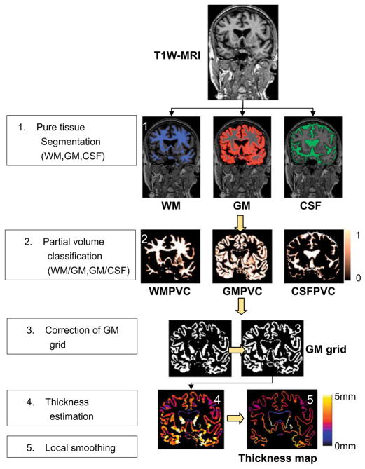
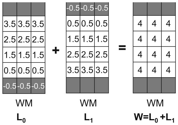
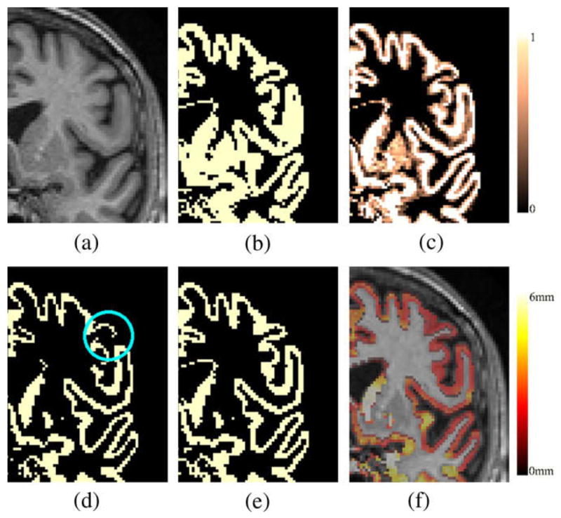
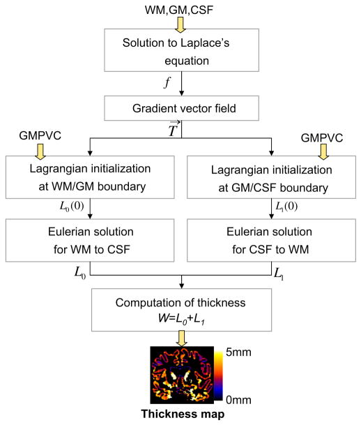
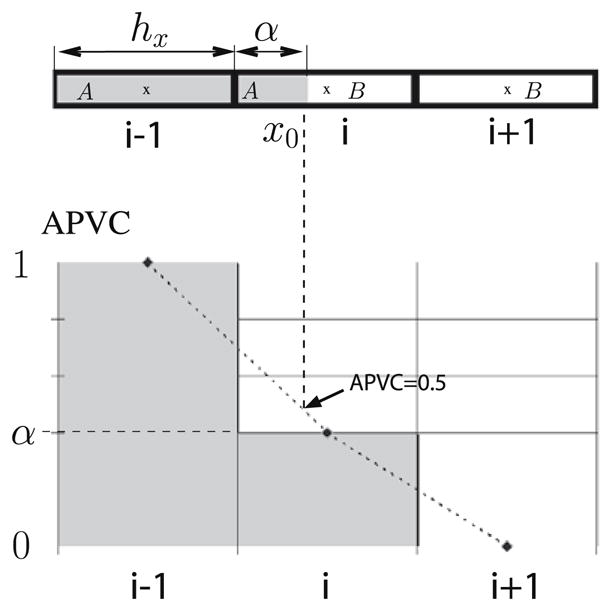
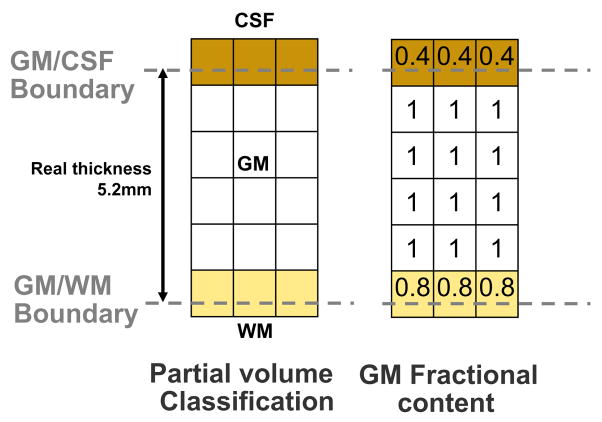
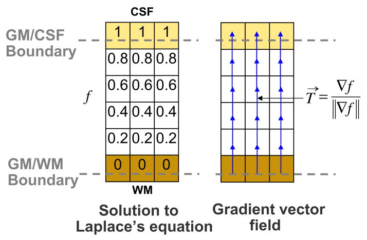
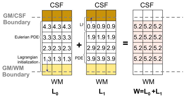
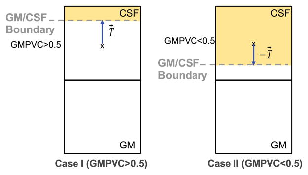
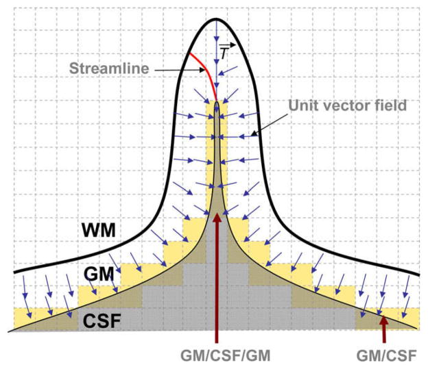
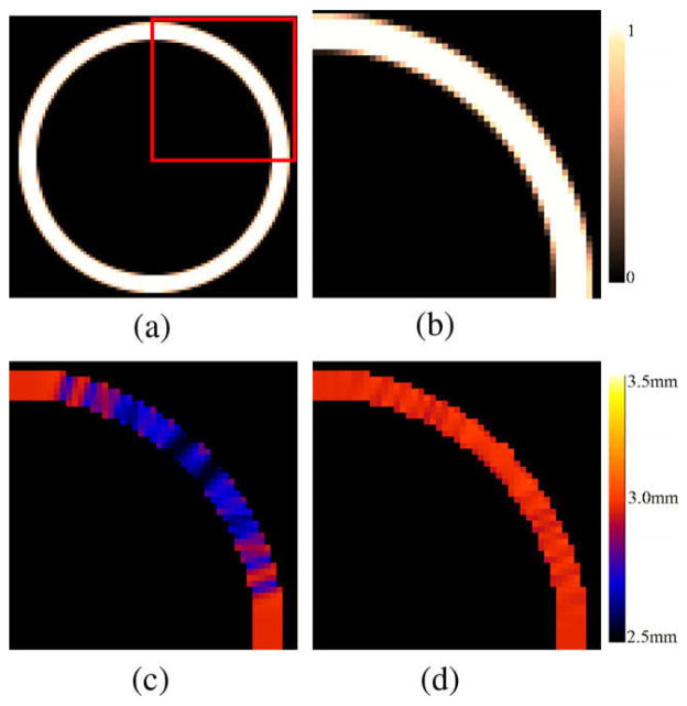
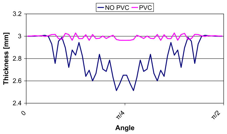
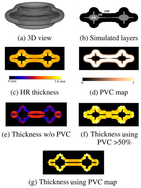
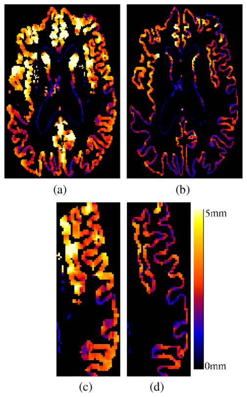
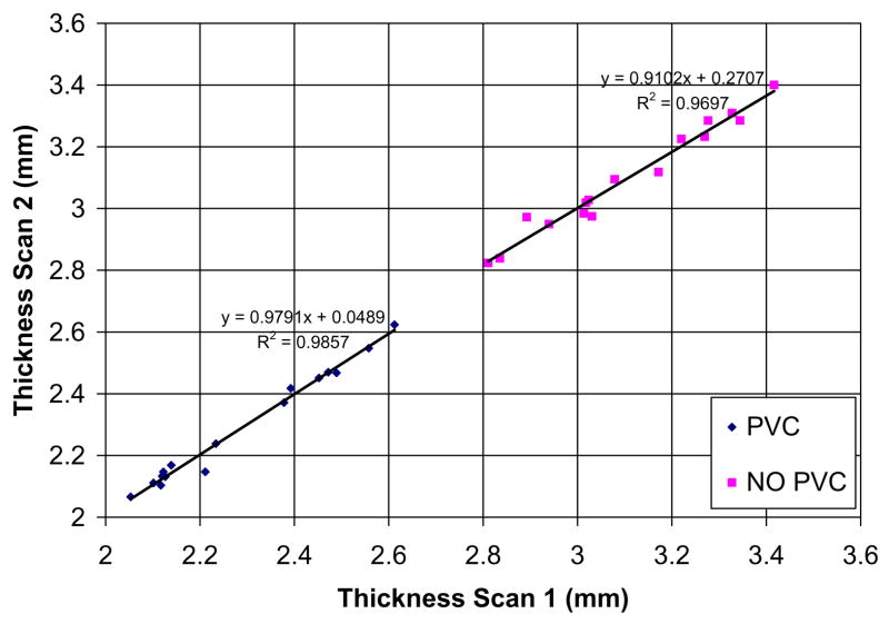
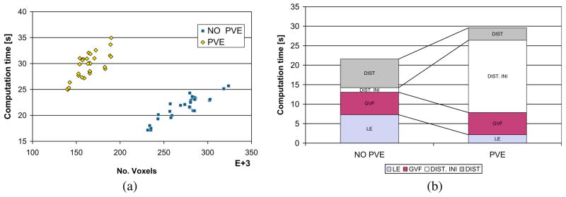
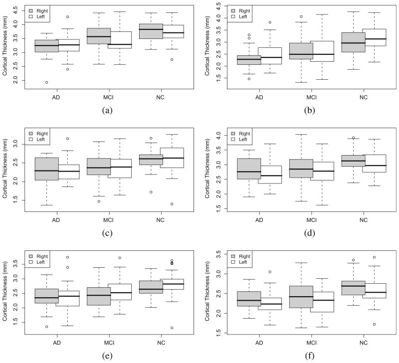
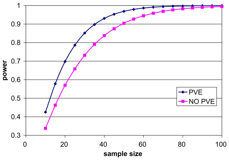
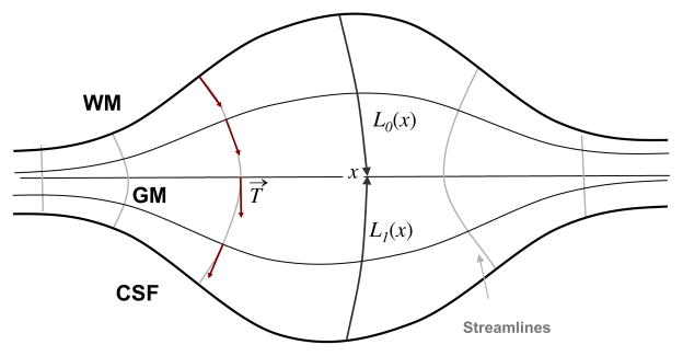
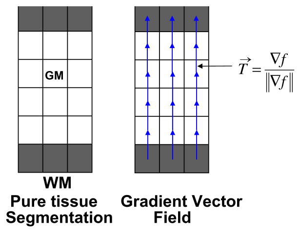
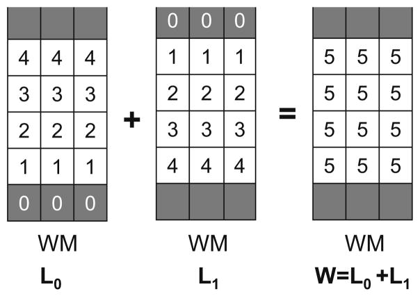
Accurate cortical thickness estimation is important for the study of many neurodegenerative diseases. Many approaches have been previously proposed, which can be broadly categorised as mesh-based and voxel-based. While the mesh-based approaches can potentially achieve subvoxel resolution, they usually lack the computational efficiency needed for clinical applications and large database studies. In contrast, voxel-based approaches, are computationally efficient, but lack accuracy. The aim of this paper is to propose a novel voxel-based method based upon the Laplacian definition of thickness that is both accurate and computationally efficient. A framework was developed to estimate and integrate the partial volume information within the thickness estimation process. Firstly, in a Lagrangian step, the boundaries are initialized using the partial volume information. Subsequently, in an Eulerian step, a pair of partial differential equations are solved on the remaining voxels to finally compute the thickness. Using partial volume information significantly improved the accuracy of the thickness estimation on synthetic phantoms, and improved reproducibility on real data. Significant differences in the hippocampus and temporal lobe between healthy controls (NC), mild cognitive impaired (MCI) and Alzheimer's disease (AD) patients were found on clinical data from the ADNI database. We compared our method in terms of precision, computational speed and statistical power against the Eulerian approach. With a slight increase in computation time, accuracy and precision were greatly improved. Power analysis demonstrated the ability of our method to yield statistically significant results when comparing AD and NC. Overall, with our method the number of samples is reduced by 25% to find significant differences between the two groups.

摘要

准确估计皮质厚度对于许多神经退行性疾病的研究至关重要。此前已经提出了许多方法,大致可分为基于网格和基于体素的方法。虽然基于网格的方法有可能实现亚体素分辨率,但它们通常缺乏临床应用和大型数据库研究所需的计算效率。相比之下,基于体素的方法计算效率高,但缺乏准确性。本文的目的是提出一种基于厚度拉普拉斯定义的新颖的基于体素的方法,该方法既准确又计算高效。开发了一个框架来估计和整合厚度估计过程中的部分体积信息。首先,在拉格朗日步骤中,使用部分体积信息初始化边界。随后,在欧拉步骤中,在剩余的体素上求解一对偏微分方程,最终计算厚度。使用部分体积信息显著提高了合成体模上厚度估计的准确性,并提高了真实数据的可重复性。在阿尔茨海默病神经成像计划(ADNI)数据库的临床数据中,发现健康对照(NC)、轻度认知障碍(MCI)和阿尔茨海默病(AD)患者的海马体和颞叶存在显著差异。我们将我们的方法在精度、计算速度和统计功效方面与欧拉方法进行了比较。在计算时间略有增加的情况下,准确性和精度得到了极大提高。功效分析表明,在比较AD和NC时,我们的方法能够产生具有统计学意义的结果。总体而言,使用我们的方法,样本数量减少了25%,以发现两组之间的显著差异。

https://cdn.ncbi.nlm.nih.gov/pmc/blobs/a41b/3068613/6d6d346b9435/nihms238471f24.jpg
https://cdn.ncbi.nlm.nih.gov/pmc/blobs/a41b/3068613/1804fb491830/nihms238471f1.jpg
https://cdn.ncbi.nlm.nih.gov/pmc/blobs/a41b/3068613/355d0c60f60a/nihms238471f2.jpg
https://cdn.ncbi.nlm.nih.gov/pmc/blobs/a41b/3068613/1bf39e255495/nihms238471f3.jpg
https://cdn.ncbi.nlm.nih.gov/pmc/blobs/a41b/3068613/29952d379f68/nihms238471f4.jpg
https://cdn.ncbi.nlm.nih.gov/pmc/blobs/a41b/3068613/71909a9afa55/nihms238471f5.jpg
https://cdn.ncbi.nlm.nih.gov/pmc/blobs/a41b/3068613/69a16918c3d1/nihms238471f6.jpg
https://cdn.ncbi.nlm.nih.gov/pmc/blobs/a41b/3068613/cc6d82a54542/nihms238471f7.jpg
https://cdn.ncbi.nlm.nih.gov/pmc/blobs/a41b/3068613/ffca2d19a152/nihms238471f8.jpg
https://cdn.ncbi.nlm.nih.gov/pmc/blobs/a41b/3068613/11b773b96b92/nihms238471f9.jpg
https://cdn.ncbi.nlm.nih.gov/pmc/blobs/a41b/3068613/1434fa9023a3/nihms238471f10.jpg
https://cdn.ncbi.nlm.nih.gov/pmc/blobs/a41b/3068613/42c9b353a96c/nihms238471f11.jpg
https://cdn.ncbi.nlm.nih.gov/pmc/blobs/a41b/3068613/e54a9dd772e2/nihms238471f12.jpg
https://cdn.ncbi.nlm.nih.gov/pmc/blobs/a41b/3068613/75048430438b/nihms238471f13.jpg
https://cdn.ncbi.nlm.nih.gov/pmc/blobs/a41b/3068613/2cef6e6fc8e4/nihms238471f14.jpg
https://cdn.ncbi.nlm.nih.gov/pmc/blobs/a41b/3068613/fc1f3cbacf7d/nihms238471f15.jpg
https://cdn.ncbi.nlm.nih.gov/pmc/blobs/a41b/3068613/b85559c5d000/nihms238471f16.jpg
https://cdn.ncbi.nlm.nih.gov/pmc/blobs/a41b/3068613/ff3fa7653176/nihms238471f17.jpg
https://cdn.ncbi.nlm.nih.gov/pmc/blobs/a41b/3068613/9a2d7dbff507/nihms238471f18.jpg
https://cdn.ncbi.nlm.nih.gov/pmc/blobs/a41b/3068613/675950180571/nihms238471f19.jpg
https://cdn.ncbi.nlm.nih.gov/pmc/blobs/a41b/3068613/1c38c37c1264/nihms238471f20.jpg
https://cdn.ncbi.nlm.nih.gov/pmc/blobs/a41b/3068613/54d4921ad4bd/nihms238471f21.jpg
https://cdn.ncbi.nlm.nih.gov/pmc/blobs/a41b/3068613/1dc609eaea21/nihms238471f22.jpg
https://cdn.ncbi.nlm.nih.gov/pmc/blobs/a41b/3068613/902cd9edbb9c/nihms238471f23.jpg
https://cdn.ncbi.nlm.nih.gov/pmc/blobs/a41b/3068613/6d6d346b9435/nihms238471f24.jpg

相似文献

1
Automated voxel-based 3D cortical thickness measurement in a combined Lagrangian-Eulerian PDE approach using partial volume maps.在使用部分体积图的拉格朗日 - 欧拉偏微分方程组合方法中基于体素的自动三维皮质厚度测量。
Med Image Anal. 2009 Oct;13(5):730-43. doi: 10.1016/j.media.2009.07.003. Epub 2009 Jul 10.
2
Automatic delineation of sulci and improved partial volume classification for accurate 3D voxel-based cortical thickness estimation from MR.自动勾勒脑沟并改进部分容积分类,以从磁共振成像中进行准确的基于体素的三维皮质厚度估计。
Med Image Comput Comput Assist Interv. 2008;11(Pt 1):253-61. doi: 10.1007/978-3-540-85988-8_31.
3
A hybrid Eulerian-Lagrangian approach for thickness, correspondence, and gridding of annular tissues.一种用于环形组织厚度、对应关系和网格化的混合欧拉-拉格朗日方法。
IEEE Trans Image Process. 2007 Mar;16(3):636-48. doi: 10.1109/tip.2007.891072.
4
Hippocampal volume change measurement: quantitative assessment of the reproducibility of expert manual outlining and the automated methods FreeSurfer and FIRST.海马体积变化测量:专家手动勾勒以及自动化方法FreeSurfer和FIRST的可重复性定量评估。
Neuroimage. 2014 May 15;92:169-81. doi: 10.1016/j.neuroimage.2014.01.058. Epub 2014 Feb 9.
5
Validation of a fully automated 3D hippocampal segmentation method using subjects with Alzheimer's disease mild cognitive impairment, and elderly controls.使用患有阿尔茨海默病轻度认知障碍的受试者和老年对照对一种全自动三维海马体分割方法进行验证。
Neuroimage. 2008 Oct 15;43(1):59-68. doi: 10.1016/j.neuroimage.2008.07.003. Epub 2008 Jul 16.
6
Sulcal morphology changes and their relationship with cortical thickness and gyral white matter volume in mild cognitive impairment and Alzheimer's disease.轻度认知障碍和阿尔茨海默病中脑沟形态变化及其与皮质厚度和脑回白质体积的关系。
Neuroimage. 2008 Oct 15;43(1):103-13. doi: 10.1016/j.neuroimage.2008.07.016. Epub 2008 Jul 22.
7
A novel cortical thickness estimation method based on volumetric Laplace-Beltrami operator and heat kernel.一种基于体积拉普拉斯 - 贝尔特拉米算子和热核的新型皮质厚度估计方法。
Med Image Anal. 2015 May;22(1):1-20. doi: 10.1016/j.media.2015.01.005. Epub 2015 Feb 3.
8
Locally linear embedding (LLE) for MRI based Alzheimer's disease classification.基于磁共振成像的阿尔茨海默病分类的局部线性嵌入(LLE)
Neuroimage. 2013 Dec;83:148-57. doi: 10.1016/j.neuroimage.2013.06.033. Epub 2013 Jun 21.
9
Improved maximum a posteriori cortical segmentation by iterative relaxation of priors.通过先验的迭代松弛改进最大后验概率皮质分割
Med Image Comput Comput Assist Interv. 2009;12(Pt 2):441-9. doi: 10.1007/978-3-642-04271-3_54.
10
Baseline and longitudinal patterns of brain atrophy in MCI patients, and their use in prediction of short-term conversion to AD: results from ADNI.轻度认知障碍(MCI)患者脑萎缩的基线和纵向模式及其在预测短期转化为阿尔茨海默病(AD)中的应用:阿尔茨海默病神经影像学计划(ADNI)的结果
Neuroimage. 2009 Feb 15;44(4):1415-22. doi: 10.1016/j.neuroimage.2008.10.031. Epub 2008 Nov 5.

引用本文的文献

1
The impact of atlas-based MR attenuation correction on the diagnosis of FDG-PET/MR for Alzheimer's diseases- A simulation study combining multi-center data and ADNI-data.基于图谱的磁共振衰减校正对阿尔茨海默病 FDG-PET/MR 诊断的影响——结合多中心数据和 ADNI 数据的模拟研究。
PLoS One. 2020 Jun 3;15(6):e0233886. doi: 10.1371/journal.pone.0233886. eCollection 2020.
2
International Mind, Activities and Urban Places (iMAP) study: methods of a cohort study on environmental and lifestyle influences on brain and cognitive health.国际思维、活动与城市空间(iMAP)研究:一项关于环境和生活方式对大脑和认知健康影响的队列研究方法。
BMJ Open. 2020 Mar 18;10(3):e036607. doi: 10.1136/bmjopen-2019-036607.
3

本文引用的文献

1
Voxel-based cortical thickness measurements in MRI.磁共振成像中基于体素的皮质厚度测量。
Neuroimage. 2008 May 1;40(4):1701-10. doi: 10.1016/j.neuroimage.2008.01.027. Epub 2008 Feb 1.
2
Quantification of MR brain images by mixture density and partial volume modeling.基于混合密度和部分体积模型对磁共振脑图像进行定量分析。
IEEE Trans Med Imaging. 1993;12(3):566-74. doi: 10.1109/42.241885.
3
G*Power 3: a flexible statistical power analysis program for the social, behavioral, and biomedical sciences.G*Power 3:一款适用于社会科学、行为科学和生物医学科学的灵活的统计功效分析程序。
Stereoelectroencephalography electrode placement: Detection of blood vessel conflicts.
立体脑电图电极放置:血管冲突的检测。
Epilepsia. 2019 Sep;60(9):1942-1948. doi: 10.1111/epi.16294. Epub 2019 Jul 22.
4
Using the Anisotropic Laplace Equation to Compute Cortical Thickness.使用各向异性拉普拉斯方程计算皮质厚度。
Med Image Comput Comput Assist Interv. 2018 Sep;11072:549-556. doi: 10.1007/978-3-030-00931-1_63. Epub 2018 Sep 13.
5
An efficient algorithm for estimating brain covariance networks.一种有效的大脑协方差网络估计算法。
PLoS One. 2018 Jul 12;13(7):e0198583. doi: 10.1371/journal.pone.0198583. eCollection 2018.
6
Grey and White Matter Alterations in Juvenile Myoclonic Epilepsy: A Comprehensive Review.青少年肌阵挛癫痫中的灰质和白质改变:一项综合综述。
J Epilepsy Res. 2017 Dec 31;7(2):77-88. doi: 10.14581/jer.17013. eCollection 2017 Dec.
7
A new method to determine cortical bone thickness in CT images using a hybrid approach of parametric profile representation and local adaptive thresholds: Accuracy results.一种使用参数化轮廓表示和局部自适应阈值的混合方法在CT图像中确定皮质骨厚度的新方法:准确性结果。
PLoS One. 2017 Nov 6;12(11):e0187097. doi: 10.1371/journal.pone.0187097. eCollection 2017.
8
Partial volume model for brain MRI scan using MP2RAGE.基于 MP2RAGE 的脑 MRI 扫描部分容积模型。
Hum Brain Mapp. 2017 Oct;38(10):5115-5127. doi: 10.1002/hbm.23719. Epub 2017 Jul 5.
9
An adaptive grid for graph-based segmentation in retinal OCT.用于视网膜光学相干断层扫描中基于图的分割的自适应网格。
Proc SPIE Int Soc Opt Eng. 2014;9034. doi: 10.1117/12.2043040.
10
Heritability and genetic correlation between the cerebral cortex and associated white matter connections.大脑皮层与相关白质连接之间的遗传力和遗传相关性。
Hum Brain Mapp. 2016 Jun;37(6):2331-47. doi: 10.1002/hbm.23177. Epub 2016 Mar 23.
Behav Res Methods. 2007 May;39(2):175-91. doi: 10.3758/bf03193146.
4
A hybrid Eulerian-Lagrangian approach for thickness, correspondence, and gridding of annular tissues.一种用于环形组织厚度、对应关系和网格化的混合欧拉-拉格朗日方法。
IEEE Trans Image Process. 2007 Mar;16(3):636-48. doi: 10.1109/tip.2007.891072.
5
Spatial patterns of cortical thinning in mild cognitive impairment and Alzheimer's disease.轻度认知障碍和阿尔茨海默病中皮质变薄的空间模式。
Brain. 2006 Nov;129(Pt 11):2885-93. doi: 10.1093/brain/awl256. Epub 2006 Sep 28.
6
Measurement of cortical thickness in 3D brain MRI data: validation of the Laplacian method.三维脑部磁共振成像数据中皮质厚度的测量:拉普拉斯方法的验证
J Neuroimaging. 2006 Apr;16(2):146-53. doi: 10.1111/j.1552-6569.2006.00036.x.
7
A novel quantitative cross-validation of different cortical surface reconstruction algorithms using MRI phantom.一种使用MRI体模对不同皮质表面重建算法进行的新型定量交叉验证。
Neuroimage. 2006 Jun;31(2):572-84. doi: 10.1016/j.neuroimage.2005.12.044. Epub 2006 Feb 24.
8
Automated 3-D extraction and evaluation of the inner and outer cortical surfaces using a Laplacian map and partial volume effect classification.使用拉普拉斯映射和部分容积效应分类对内外皮质表面进行自动三维提取和评估。
Neuroimage. 2005 Aug 1;27(1):210-21. doi: 10.1016/j.neuroimage.2005.03.036.
9
Cortical thickness analysis examined through power analysis and a population simulation.通过功效分析和总体模拟对皮质厚度进行分析。
Neuroimage. 2005 Jan 1;24(1):163-73. doi: 10.1016/j.neuroimage.2004.07.045.
10
Focal decline of cortical thickness in Alzheimer's disease identified by computational neuroanatomy.通过计算神经解剖学确定的阿尔茨海默病患者皮质厚度的局灶性下降。
Cereb Cortex. 2005 Jul;15(7):995-1001. doi: 10.1093/cercor/bhh200. Epub 2004 Nov 10.