Child Psychiatry Branch, National Institute of Mental Health/NIH, Bethesda, MD 20892, USA.
Neuroimage. 2010 Jan 1;49(1):63-70. doi: 10.1016/j.neuroimage.2009.08.016. Epub 2009 Aug 13.
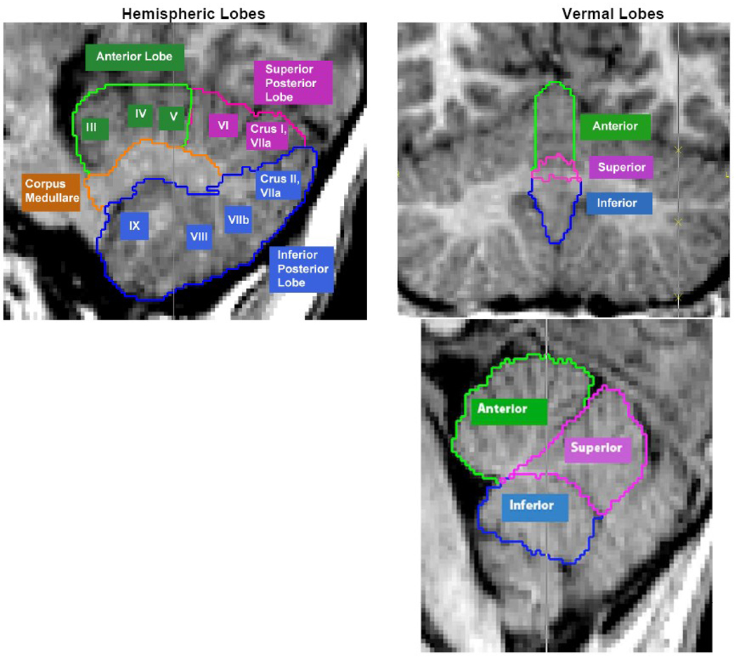
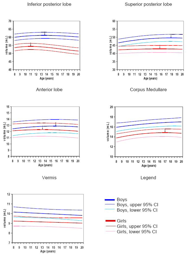
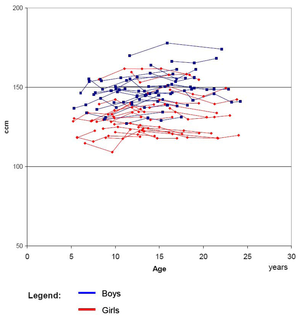
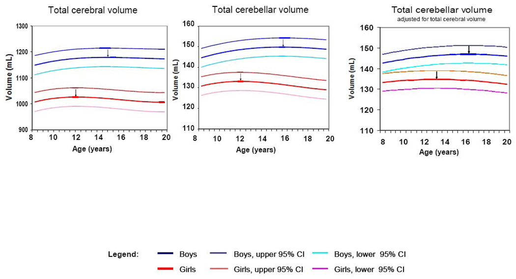
In addition to its well-established role in balance, coordination, and other motor skills, the cerebellum is increasingly recognized as a prominent contributor to a wide array of cognitive and emotional functions. Many of these capacities undergo dramatic changes during childhood and adolescence. However, accurate characterization of co-occurring anatomical changes has been hindered by lack of longitudinal data and methodologic challenges in quantifying subdivisions of the cerebellum. In this study we apply an innovative image analysis technique to quantify total cerebellar volume and 11 subdivisions (i.e. anterior, superior posterior, and inferior posterior lobes, corpus medullare, and three vermal regions) from anatomic brain MRI scans from 25 healthy females and 25 healthy males aged 5-24 years, each of whom was scanned at least three times at approximately 2-year intervals. Total cerebellum volume followed an inverted U shaped developmental trajectory peaking at age 11.8 years in females and 15.6 years in males. Cerebellar volume was 10% to 13% larger in males depending on the age of comparison and the sexual dimorphism remained significant after covarying for total brain volume. Subdivisions of the cerebellum had distinctive developmental trajectories with more phylogenetically recent regions maturing particularly late. The cerebellum's unique protracted developmental trajectories, sexual dimorphism, preferential vulnerability to environmental influences, and frequent implication in childhood onset disorders such as autism and ADHD make it a prime target for pediatric neuroimaging investigations.
除了在平衡、协调和其他运动技能方面的成熟作用外,小脑越来越被认为是广泛的认知和情感功能的重要贡献者。这些能力中的许多在儿童和青少年时期经历了巨大的变化。然而,由于缺乏纵向数据和量化小脑细分的方法学挑战,对同时发生的解剖变化的准确描述受到了阻碍。在这项研究中,我们应用了一种创新的图像分析技术,从 25 名健康女性和 25 名健康男性的解剖大脑 MRI 扫描中量化总小脑体积和 11 个细分区域(即前叶、上后叶和下后叶、髓质和三个蚓部区域),他们每个人都在大约 2 年的时间间隔内至少扫描了 3 次。总小脑体积呈倒 U 形发育轨迹,在女性中在 11.8 岁时达到峰值,在男性中在 15.6 岁时达到峰值。小脑体积在男性中比女性大 10%至 13%,具体取决于比较的年龄,并且在考虑总脑体积后,性别二态性仍然显著。小脑的细分区域具有独特的发育轨迹,其中进化上较新的区域成熟得特别晚。小脑独特的延长发育轨迹、性别二态性、对环境影响的优先脆弱性以及在儿童期发病障碍(如自闭症和 ADHD)中的频繁牵连,使其成为儿科神经影像学研究的主要目标。