Roberts D L, Williams K J, Cowen R L, Barathova M, Eustace A J, Brittain-Dissont S, Tilby M J, Pearson D G, Ottley C J, Stratford I J, Dive C
Paterson Institute for Cancer Research, University of Manchester, Manchester, UK.
Br J Cancer. 2009 Oct 20;101(8):1290-7. doi: 10.1038/sj.bjc.6605311. Epub 2009 Sep 15.
Hypoxia is as an indicator of poor treatment outcome. Consistently, hypoxic HCT116 colorectal cancer cells are resistant to oxaliplatin, although the mechanistic basis is unclear. This study sought to investigate the relative contribution of HIF-1 (hypoxia-inducible factor-1)-mediated gene expression and drug penetrance to oxaliplatin resistance using three-dimensional spheroids.
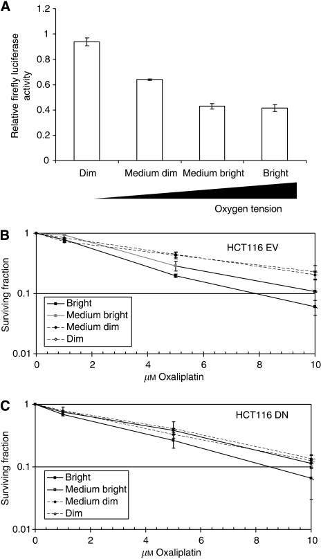
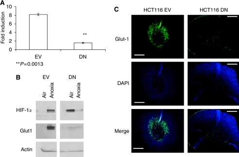
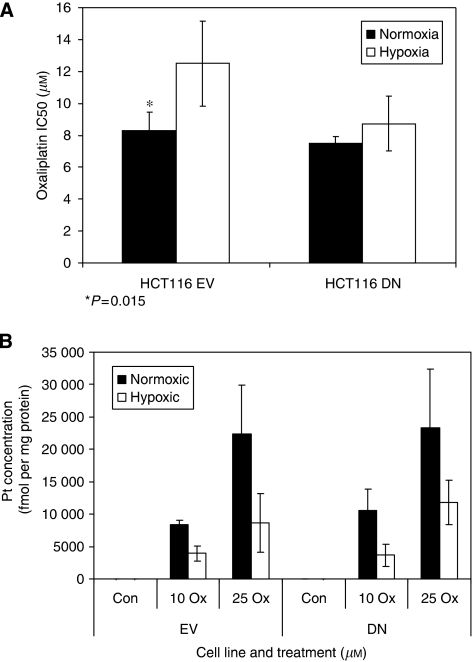
Hypoxia-inducible factor-1alpha function was suppressed by the stable expression of a dominant-negative form in HCT116 cells (DN). Cells were drug exposed as monolayer or multicellular spheroid cultures. Cells residing at differing oxygenation status were isolated from Hoechst 33342-treated spheroids using flow cytometry. Sub-populations were subjected to clonogenic survival assays and to Inductively-Coupled Plasma Mass Spectroscopy to determine oxaliplatin uptake.
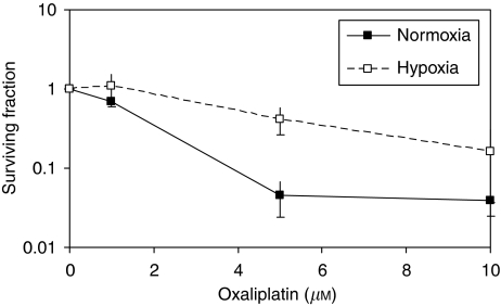
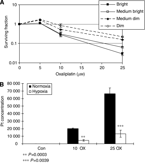
In spheroids, a sensitivity gradient (hypoxic<aerobic) was revealed by survival assays and this correlated with levels of platinum-bound DNA. The resistance of hypoxic sub-populations exceeded relative changes in adduct levels, implicating factors other than drug penetrance in cell response. Dominant-negative monolayer cells showed no resistance to oxaliplatin in hypoxia and spheroids; the relative resistance of hypoxic compared with aerobic sub-populations was reduced compared with those from controls.
Overall, data show that drug penetration, DNA damage levels and HIF-1-dependent processes, all contribute to the resistance of hypoxic cells to oxaliplatin.
缺氧是治疗效果不佳的一个指标。一直以来,缺氧的HCT116结肠癌细胞对奥沙利铂耐药,但其机制尚不清楚。本研究旨在使用三维球体研究缺氧诱导因子-1(HIF-1)介导的基因表达和药物渗透性对奥沙利铂耐药性的相对贡献。
通过在HCT116细胞(DN)中稳定表达显性阴性形式来抑制缺氧诱导因子-1α的功能。细胞作为单层或多细胞球体培养物进行药物处理。使用流式细胞术从经Hoechst 33342处理的球体中分离处于不同氧合状态的细胞。对亚群进行克隆存活分析和电感耦合等离子体质谱分析以确定奥沙利铂的摄取。
在球体中,存活分析显示出敏感性梯度(缺氧<需氧),这与铂结合DNA的水平相关。缺氧亚群的耐药性超过了加合物水平的相对变化,这表明除了药物渗透性之外,还有其他因素参与细胞反应。显性阴性单层细胞在缺氧和球体中对奥沙利铂没有耐药性;与对照组相比,缺氧亚群与需氧亚群相比的相对耐药性降低。
总体而言,数据表明药物渗透性、DNA损伤水平和HIF-1依赖性过程均导致缺氧细胞对奥沙利铂产生耐药性。