Murashima Yoshiya L, Suzuki Jiro, Yoshii Mitsunobu
Division of Psychobiology, Tokyo Institute of Psychiatry, Tokyo, Japan.
Gene Regul Syst Bio. 2008 Aug 27;2:267-74.
Epileptic mutant EL mice show secondary generalized seizures. Seizure discharges initiate in the parietal cortex and generalize through the hippocampus. We have previously demonstrated an increase in the activity of inducible nitric oxide synthetase (iNOS) as well as a decrease in the activity of superoxide dismutase (SOD) in the hippocampus of EL mice, suggesting that cell toxic free radicals are increased in the brain of EL mice. In parallel with this, neurotrophic factors were significantly increased in the hippocampus of EL mice in earlier developmental stages before exhibiting frequent seizures. These findings were no longer present after frequent seizures, suggesting that these events may trigger ictogenesis. On the other hand, it is reported that limbic seizures rapidly induce cytokines and related inflammatory mediators. It remains to be seen, however, whether cytokines contribute to the transition from interictal to ictal state. The present study was designed to address this issue using EL mice.
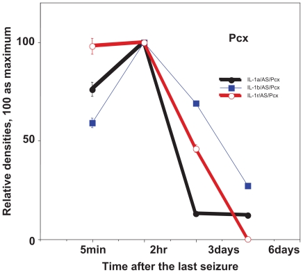
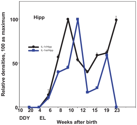
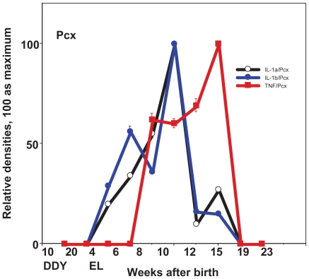
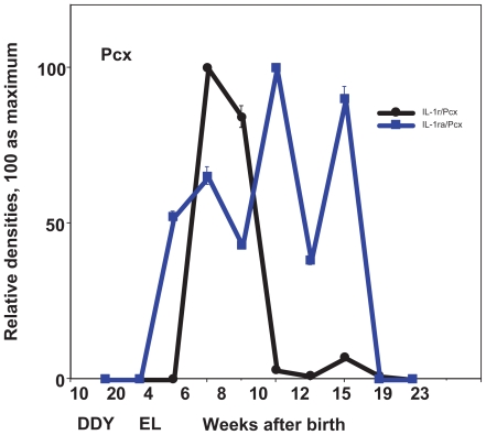
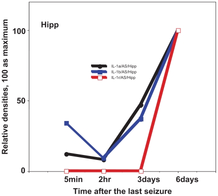
EL mice at the age from 4 to 23 weeks and their control animal, DDY mice at the age of 10 and 20 weeks were used. Seizures were induced in EL mice once every week since 5 weeks. Cytokines, such as interleukin-1 alpha (IL-1a), interleukin 1-beta (IL-1b), IL-6, IL-1 receptor (IL-1r), IL-1 receptor antagonist (IL-ra) and tumor necrosis factor alpha (TNF-a) were examined by Western blotting in the 'focus complex' of brain (namely, in the parietal cortex and hippocampus) of EL mice in the interictal period at different developmental stages. In 15 week old EL mice, which show seizures once a week, these cytokines were similarly determined 5 min, 2 hr, 4 hr, 11 hr, 24 hr, 3 days and 6 days after the last seizure induced.
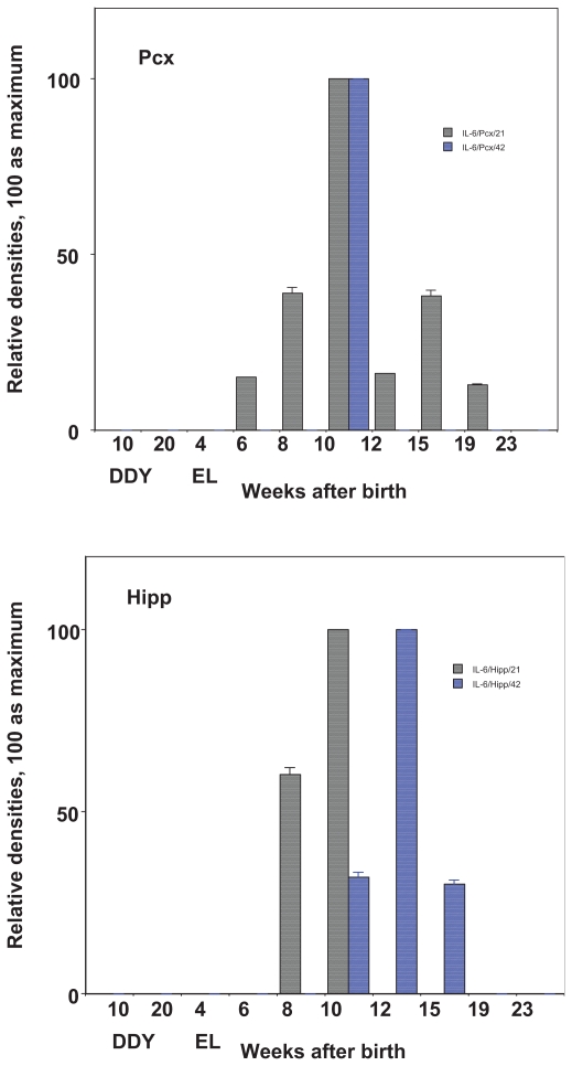
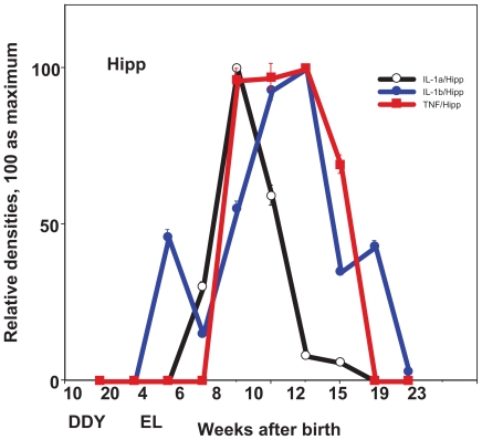
A significant increase in the level of cytokines was observed in the brain of EL mice at any stages during development, compared with the level of cytokines in the brain of control DDY. Cytokines were increased predominantly before experiencing frequent seizures. In EL mice at the age of 15 weeks, the level of cytokines in the hippocampus was highest 6 days after seizures. In the parietal cortex, cytokines were most highly expressed 2 hr after seizures. The results indicate that cytokines were kept up-regulated until next seizures in the hippocampus, whereas they were transiently up-regulated immediately after seizures in the parietal cortex.
It is concluded that in the brain of EL mice, pro-inflammatory cytokines are increased progressively and periodically in association with the development and the seizure activity, respectively. A periodic increase of cytokines prior to the next seizure episode may play a role in triggering the ictal activity. Namely, alteration of region-specific cytokines may induce ictal activities from the interictal state. It is conceivable that inflammatory cytokines may work together with neuronal factors during epileptogenesis and in the transition from interictal to ictal state.
癫痫突变型EL小鼠表现出继发性全身性癫痫发作。癫痫放电起始于顶叶皮质,并通过海马体扩散。我们之前已经证明,EL小鼠海马体中诱导型一氧化氮合酶(iNOS)的活性增加,同时超氧化物歧化酶(SOD)的活性降低,这表明EL小鼠大脑中的细胞毒性自由基增加。与此同时,在EL小鼠出现频繁癫痫发作之前的早期发育阶段,海马体中的神经营养因子显著增加。频繁癫痫发作后这些发现不再存在,这表明这些事件可能触发癫痫发作的产生。另一方面,据报道边缘性癫痫发作会迅速诱导细胞因子和相关炎症介质。然而,细胞因子是否促成从发作间期到发作期的转变仍有待观察。本研究旨在使用EL小鼠解决这个问题。
使用4至23周龄的EL小鼠及其对照动物,10周龄和20周龄的DDY小鼠。自5周龄起,每周对EL小鼠诱导一次癫痫发作。通过蛋白质印迹法检测不同发育阶段发作间期EL小鼠大脑“病灶复合体”(即顶叶皮质和海马体)中白细胞介素-1α(IL-1α)、白细胞介素1-β(IL-1β)、IL-6、IL-1受体(IL-1r)、IL-1受体拮抗剂(IL-ra)和肿瘤坏死因子α(TNF-α)等细胞因子。在每周发作一次癫痫的15周龄EL小鼠中,在最后一次诱导癫痫发作后的5分钟、2小时、4小时、11小时、24小时、3天和6天同样测定这些细胞因子。
与对照DDY小鼠大脑中的细胞因子水平相比,在发育的任何阶段,EL小鼠大脑中的细胞因子水平均显著升高。细胞因子主要在经历频繁癫痫发作之前增加。在15周龄的EL小鼠中,海马体中的细胞因子水平在癫痫发作后6天最高。在顶叶皮质中,细胞因子在癫痫发作后2小时表达最高。结果表明,海马体中的细胞因子在下次癫痫发作前一直上调,而在顶叶皮质中,细胞因子在癫痫发作后立即短暂上调。
得出结论,在EL小鼠大脑中,促炎细胞因子分别与发育和癫痫发作活动相关,逐渐且周期性地增加。在下一次癫痫发作前细胞因子的周期性增加可能在触发发作活动中起作用。也就是说,区域特异性细胞因子的改变可能从发作间期状态诱导发作活动。可以想象,炎症细胞因子可能在癫痫发生过程中以及从发作间期到发作期的转变中与神经元因子共同起作用。