Chowdhury Manzurul, Endo Masaki, Chiba Toshimi, Kudara Norihiko, Oana Shuhei, Sato Kunihiko, Akasaka Risaburo, Tomita Kazumitsu, Fujiwara Saori, Mizutani Tomomi, Sugai Tamotsu, Takikawa Yasuhiro, Suzuki Kazuyuki
Department of Gastroenterology and Hepatology, Iwate Medical University, Morioka, Iwate 020-8505, Japan.
Gastroenterol Res Pract. 2009;2009:835258. doi: 10.1155/2009/835258. Epub 2009 Nov 5.
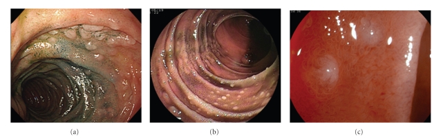
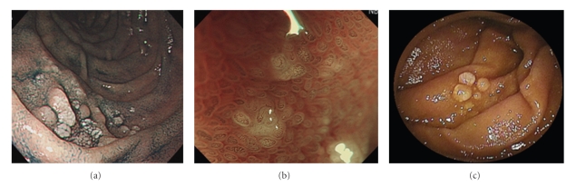
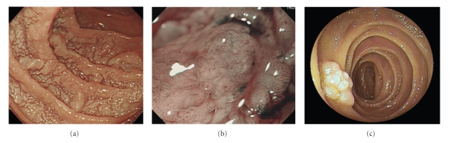
Follicular lymphomas occur rarely in the gastrointestinal tract, representing only 1-3% of all gastrointestinal tract B-cell non-Hodgkin lymphomas. We describe endoscopic analysis of 3 cases of follicular lymphoma in the small intestine using double-balloon endoscopy. Double-balloon endoscopy revealed multiple nodular lesions and elevated white patches, multiple polypoid lesions, and scattered white polypoid and nodular lesions in the duodenum and small intestine. Fuji Intelligent Chromo Endoscopy demonstrated small, whitish nodules, and narrow-band imaging showed a coiled, elongated vascular pattern within the elevated lesions. These cases are the first follicular lymphomas in the small intestine evaluated using narrow-band imaging or Fuji Intelligent Chromo Endoscopy to be reported.
滤泡性淋巴瘤很少发生于胃肠道,仅占所有胃肠道B细胞非霍奇金淋巴瘤的1%-3%。我们描述了3例小肠滤泡性淋巴瘤的双气囊内镜分析。双气囊内镜检查发现十二指肠和小肠有多个结节性病变、白色隆起斑、多个息肉样病变以及散在的白色息肉样和结节性病变。富士智能染色内镜显示有小的白色结节,窄带成像显示隆起病变内有盘绕、拉长的血管形态。这些病例是首次报道的使用窄带成像或富士智能染色内镜评估的小肠滤泡性淋巴瘤。