Wiechmann Allan F, Hollaway Lindsey R, Rada Jody A Summers
Department of Cell Biology, University of Oklahoma Health Sciences Center, Oklahoma City, OK 73126-0901, USA.
Mol Vis. 2009 Nov 17;15:2384-403.
Melatonin receptors are seven-pass G protein-coupled receptors located in many tissues throughout the body, including the corneal epithelium (CE), and relay circadian signals to the target cells. The purpose of this study was to determine more precisely the cellular distribution of the melatonin receptors in the surface cells of the CE of Xenopus laevis, and to examine the relative distribution of melatonin receptor subtype expression at different times during the circadian cycle.
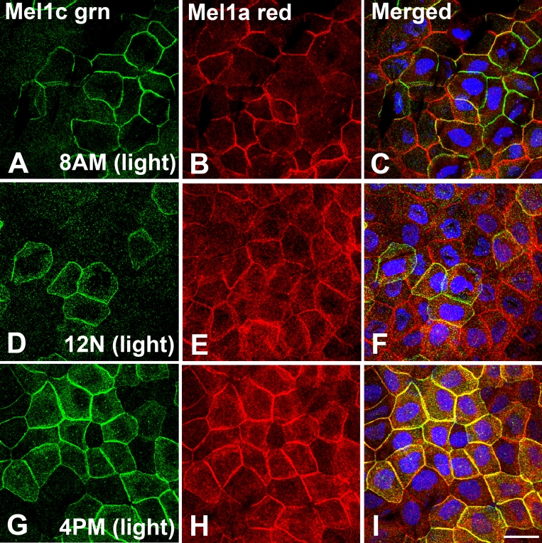
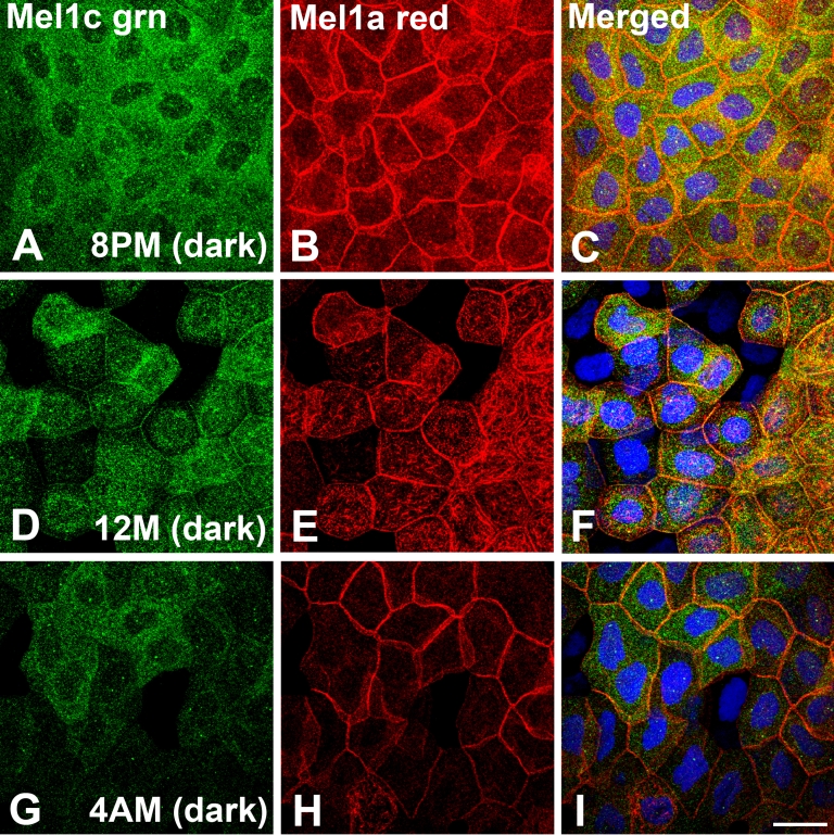
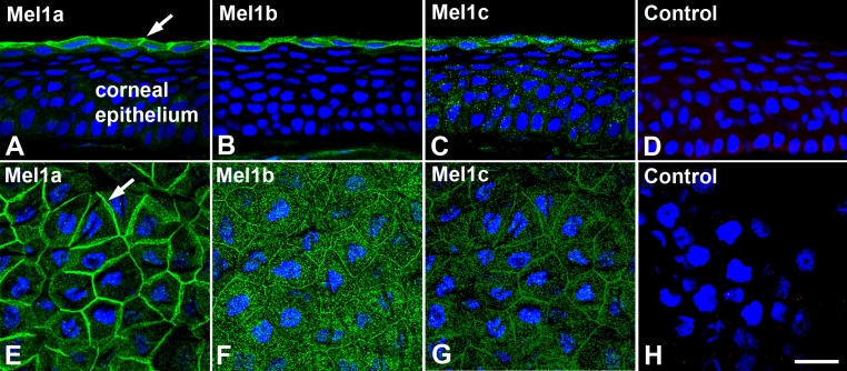
Cryostat sections and whole corneas of adult Xenopus laevis were processed for immunocytochemistry using antibodies specific for each of the three melatonin receptor subtypes (Mel1a, Mel1b, and Mel1c). For the circadian studies, corneas were obtained from euthanized frogs at 4-h intervals during a 24-h period under a 12 h:12 h light-dark cycle. Double-label immunocytochemistry was performed using a Mel1a antibody in combination with antibodies against Mel1b, Mel1c, or the zonula occludens protein ZO-1. Corneal whole-mount specimens and corneal sections were analyzed by laser-scanning confocal microscopy.
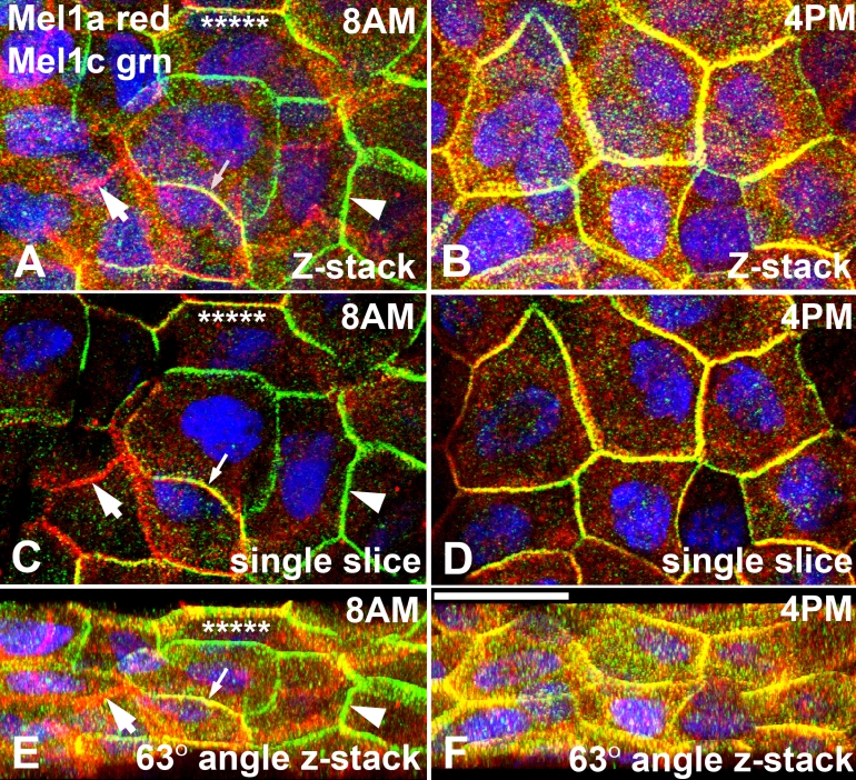
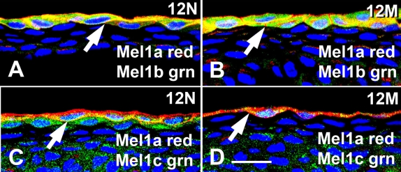
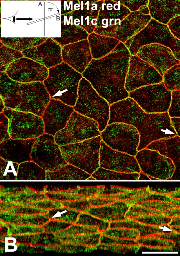
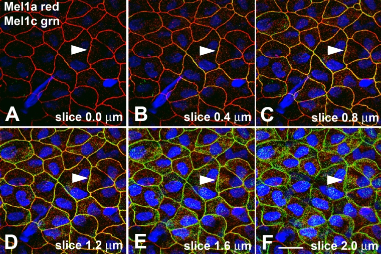
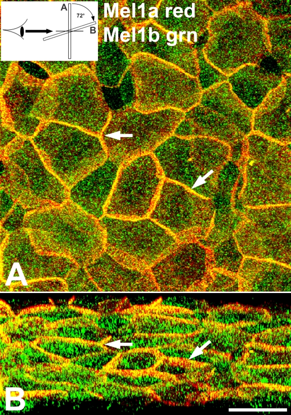
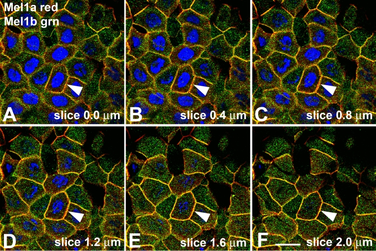
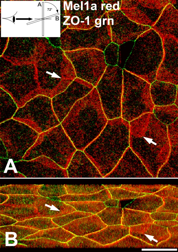
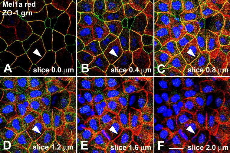
All three melatonin receptor subtypes were expressed on the surface and sub-superficial layer of CE cells, but with different sub-cellular distributions. The Mel1a receptor was highly localized to the lateral plasma membrane of the surface CE, but also displayed cytoplasmic localization at some times of day, especially at night. Mel1c showed a similar pattern of labeling to Mel1a, but there were some distinctive differences, insofar as the Mel1c receptors were usually located immediately basal to the Mel1a receptors. The relative degree of membrane and cytoplasmic labeling of the Mel1c receptor also oscillated during the 24-h period, but was out of phase with the changes that occurred in the Mel1a receptor localization. Furthermore, in the late afternoon time point, the Mel1a and Mel1c receptors were highly co-localized, suggestive of heterodimerization, whereas at other time points, the two receptors were distinctly not co-localized. Double-label immunocytochemistry of Mel1a and ZO-1 demonstrated that the Mel1a receptor was located basal to the tight junctions, on the lateral membrane in very close proximity to the ZO-1 protein.
Mel1a, Mel1b, and Mel1c receptor subtypes are expressed in the lateral plasma membrane of the Xenopus surface CE, at a position in close proximity to the tight junctions that form the corneal diffusion barrier. The very close association of the Mel1a receptors to the ZO-1 peripheral membrane tight junction proteins is suggestive of a potential role for melatonin in influencing the rate of tight junction formation or breakdown. The transient co-localization of Mel1a and Mel1c late in the light period is suggestive of formation of heterodimers that may influence receptor responsiveness and/or activity during specific periods of the day. The dynamic daily changes in melatonin receptor subtype expression and localization in the surface CE supports the concept that melatonin signaling may affect circadian activities of the surface epithelium of the cornea.
褪黑素受体是七次跨膜的G蛋白偶联受体,存在于全身许多组织中,包括角膜上皮(CE),并将昼夜节律信号传递给靶细胞。本研究的目的是更精确地确定非洲爪蟾角膜上皮表面细胞中褪黑素受体的细胞分布,并研究昼夜节律周期不同时间点褪黑素受体亚型表达的相对分布。
使用针对三种褪黑素受体亚型(Mel1a、Mel1b和Mel1c)各自的特异性抗体,对成年非洲爪蟾的冰冻切片和全角膜进行免疫细胞化学处理。对于昼夜节律研究,在12小时光照:12小时黑暗周期的24小时内,每隔4小时从安乐死的青蛙获取角膜。使用Mel1a抗体与抗Mel1b、Mel1c或紧密连接蛋白ZO-1的抗体进行双重标记免疫细胞化学。通过激光扫描共聚焦显微镜分析角膜整装标本和角膜切片。
所有三种褪黑素受体亚型均在角膜上皮细胞的表面和浅表层表达,但具有不同的亚细胞分布。Mel1a受体高度定位于角膜上皮表面的外侧质膜,但在一天中的某些时间,尤其是夜间,也表现出细胞质定位。Mel1c显示出与Mel1a相似的标记模式,但存在一些明显差异,因为Mel1c受体通常位于Mel1a受体的紧邻基部。Mel1c受体的膜和细胞质标记相对程度在24小时内也有波动,但与Mel1a受体定位的变化不同步。此外,在下午晚些时候的时间点,Mel1a和Mel1c受体高度共定位,提示异二聚体形成,而在其他时间点,这两种受体明显不共定位。Mel1a和ZO-1的双重标记免疫细胞化学表明,Mel1a受体位于紧密连接的基部,在外侧膜上非常靠近ZO-1蛋白。
Mel1a、Mel1b和Mel1c受体亚型在非洲爪蟾角膜表面的外侧质膜中表达,位于形成角膜扩散屏障的紧密连接附近。Mel1a受体与ZO-1外周膜紧密连接蛋白的紧密关联提示褪黑素在影响紧密连接形成或分解速率方面可能发挥潜在作用。光照期后期Mel1a和Mel1c的短暂共定位提示形成了可能在一天特定时期影响受体反应性和/或活性的异二聚体。角膜上皮表面褪黑素受体亚型表达和定位的每日动态变化支持了褪黑素信号可能影响角膜表面上皮昼夜活动的概念。