Shriners Hospitals for Children, Erie, PA, USA.
Spine (Phila Pa 1976). 2009 Dec 1;34(25):2782-6. doi: 10.1097/BRS.0b013e3181c11853.
Longitudinal radiographic study of patients with progressive idiopathic scoliosis.
To determine the relative contributions of vertebral and disc wedging to the increase in Cobb angle during 3 phases of adolescent skeletal growth and maturation.
Both disc wedging and vertebral body wedging are found in progressive scoliosis, but their relative contribution to curve progression over time is unknown. Which occurs first is important for understanding how scoliosis progresses and for developing methods to halt progression. Previous studies have not properly identified maturity, and provide conflicting results.
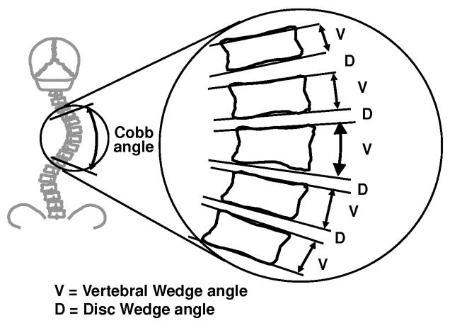
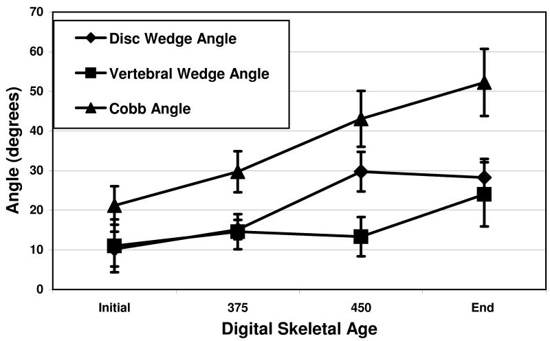
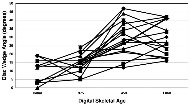
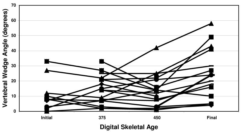
Eighteen girls were followed through their adolescent growth spurt with serial spine and hand skeletal age radiographs. Each Cobb angle was divided into disc wedge angles and vertebral wedge angles. The corresponding hand radiographs provided a measure of maturity level, the Digital Skeletal Age (DSA). The disc versus bone contributions to the Cobb angle were then compared during 3 growth phases: before the growth spurt, during the growth spurt and after the growth spurt. Significance of relative changes was assessed with the Wilcoxon 2-sided mean rank test.
Before the growth spurt, there was no difference in relative contributions of the disc and the bone (3 degrees vs. 0 degrees, P = 0.38) to curve progression. During the growth spurt, the mean disc component progressed significantly more than that of the vertebrae (15 degrees vs. 0 degrees, P = 0.0002). This reversed following the growth spurt with the vertebral component progressing more than the disc (10 degrees vs. 0 degrees, P = 0.01).
Adolescent idiopathic scoliosis initially increases through disc wedging during the rapid growth spurt with progressive vertebral wedging occurring later.
对进展性特发性脊柱侧凸患者的纵向影像学研究。
确定在青少年骨骼生长和成熟的 3 个阶段中,椎骨和椎间盘楔形变形对 Cobb 角增加的相对贡献。
进展性脊柱侧凸中既有椎间盘楔形变形,也有椎体楔形变形,但它们对随时间推移的曲线进展的相对贡献尚不清楚。这两种情况哪个先发生对于理解脊柱侧凸如何进展以及开发阻止进展的方法很重要。以前的研究没有正确识别成熟度,并且提供了相互矛盾的结果。
18 名女孩通过连续的脊柱和手部骨骼年龄 X 光片接受了她们青春期生长突增的随访。每个 Cobb 角都被分为椎间盘楔形角和椎体楔形角。相应的手部 X 光片提供了成熟度水平的测量值,即数字骨骼年龄(DSA)。然后在 3 个生长阶段比较 Cobb 角的椎间盘与骨的贡献:生长突增前、生长突增中和生长突增后。使用 Wilcoxon 双侧平均秩检验评估相对变化的显著性。
在生长突增前,椎间盘和骨骼对曲线进展的相对贡献没有差异(3 度对 0 度,P = 0.38)。在生长突增期间,椎间盘的平均成分进展明显超过椎骨(15 度对 0 度,P = 0.0002)。生长突增后这种情况发生逆转,椎骨成分的进展超过椎间盘(10 度对 0 度,P = 0.01)。
青少年特发性脊柱侧凸最初通过生长突增期间的椎间盘楔形变形增加,随后发生渐进性椎体楔形变形。