AA 1105 MCN, Vanderbilt University Institute of Imaging Science, Department of Radiology and Radiological Sciences, Vanderbilt University, Nashville, TN 37232-2310, USA.
Epilepsy Res. 2010 Feb;88(2-3):168-78. doi: 10.1016/j.eplepsyres.2009.10.018. Epub 2009 Nov 27.
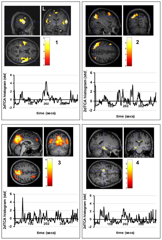
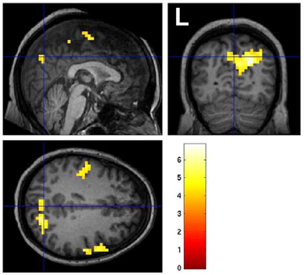
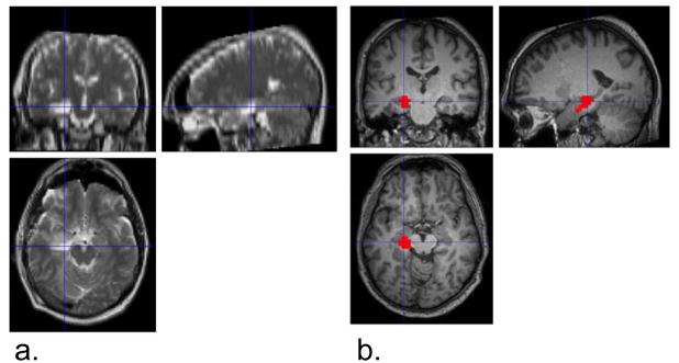
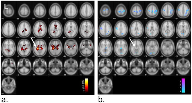
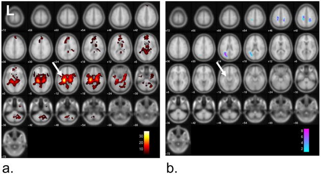
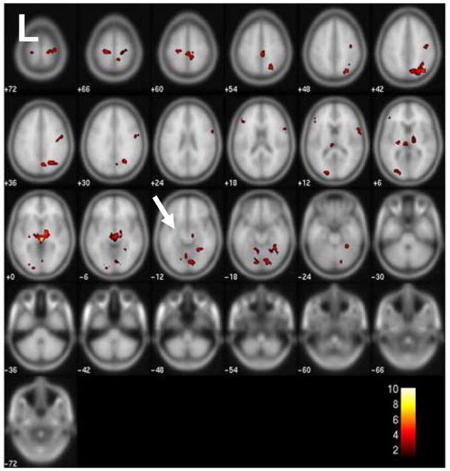
The purpose of this study was to determine transient functional signal activity in a small, homogeneous group of left temporal lobe epilepsy (TLE) patients, without the use of EEG; and to use one of these activated regions to identify a possible epileptogenic network across the whole brain in this group. Resting functional MRI scanning was performed on five left TLE patients who underwent selective amygdalohippocampectomy resulting in seizure control and 10 healthy control subjects. Activation maps of functional signal peaks were calculated using a data-driven analysis, 2dTCA, across the group of patients. In addition to the expected region of activation in the left anterior hippocampus, the results of the 2dTCA analysis revealed activity in the bilateral insular cortex and default-mode network which are not commonly reported using fMRI, but are supported by other electrical and functional changes. The region of activation corresponding to the anterior hippocampal region of resection (presumably the epileptogenic region) was used as a seed region for fMRI functional connectivity analysis. This revealed increased negative connectivity in the patients as compared to controls across a network including thalamic, brainstem, frontal and parietal regions consistent with theories of inhibited function in subcortical and cortical structures during ictal propagation.
这项研究的目的是在一小群左颞叶癫痫(TLE)患者中确定短暂的功能信号活动,而不使用 EEG;并使用其中一个激活区域来识别该组患者整个大脑中可能的致痫网络。对 5 名接受选择性杏仁核海马切除术的左 TLE 患者和 10 名健康对照进行静息功能磁共振扫描,结果显示癫痫得到控制。使用数据驱动分析(2dTCA)在患者组中计算功能信号峰的激活图。除了左前海马的预期激活区域外,2dTCA 分析的结果还显示双侧岛叶皮层和默认模式网络的活动,这些活动通常不会通过 fMRI 报告,但得到了其他电和功能变化的支持。与切除的前海马区域(推测为致痫区)相对应的激活区域被用作 fMRI 功能连接分析的种子区域。与对照组相比,患者的功能连接呈现出负相关,这表明在包括丘脑、脑干、额叶和顶叶区域的网络中存在抑制功能,这与发作传播过程中皮质下和皮质结构的抑制功能理论一致。