Department of Biology and Center for Regenerative Biology and Medicine, Indiana University-Purdue University Indianapolis, Indianapolis, IN, USA.
BMC Biol. 2009 Nov 30;7:83. doi: 10.1186/1741-7007-7-83.
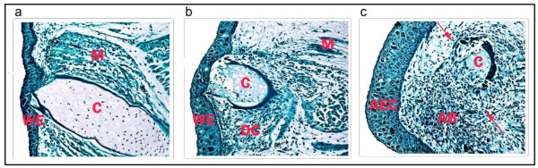
Following amputation, urodele salamander limbs reprogram somatic cells to form a blastema that self-organizes into the missing limb parts to restore the structure and function of the limb. To help understand the molecular basis of blastema formation, we used quantitative label-free liquid chromatography-mass spectrometry/mass spectrometry (LC-MS/MS)-based methods to analyze changes in the proteome that occurred 1, 4 and 7 days post amputation (dpa) through the mid-tibia/fibula of axolotl hind limbs.
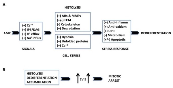
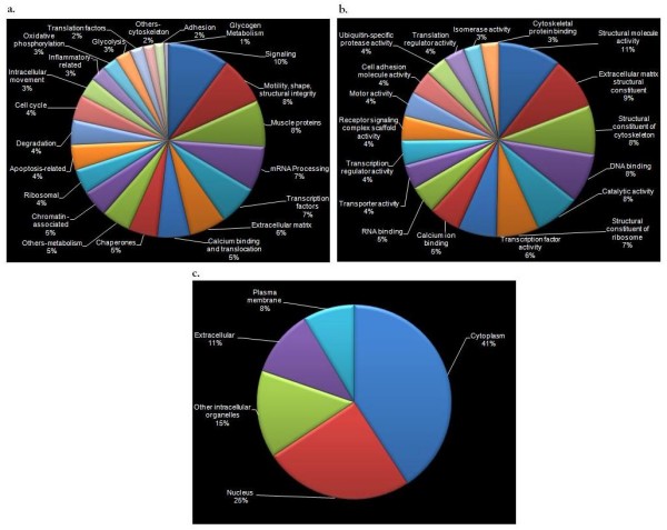
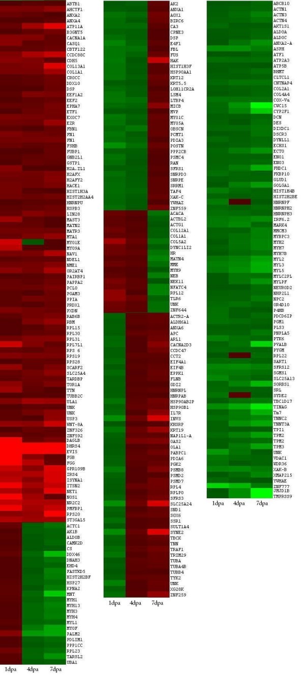
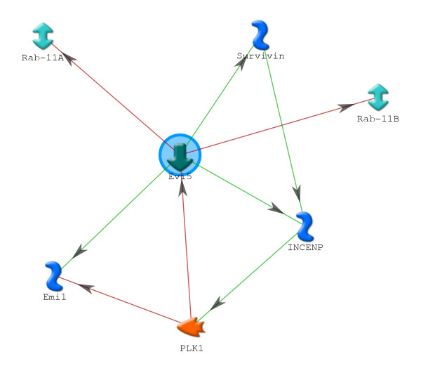
We identified 309 unique proteins with significant fold change relative to controls (0 dpa), representing 10 biological process categories: (1) signaling, (2) Ca2+ binding and translocation, (3) transcription, (4) translation, (5) cytoskeleton, (6) extracellular matrix (ECM), (7) metabolism, (8) cell protection, (9) degradation, and (10) cell cycle. In all, 43 proteins exhibited exceptionally high fold changes. Of these, the ecotropic viral integrative factor 5 (EVI5), a cell cycle-related oncoprotein that prevents cells from entering the mitotic phase of the cell cycle prematurely, was of special interest because its fold change was exceptionally high throughout blastema formation.
Our data were consistent with previous studies indicating the importance of inositol triphosphate and Ca2+ signaling in initiating the ECM and cytoskeletal remodeling characteristic of histolysis and cell dedifferentiation. In addition, the data suggested that blastema formation requires several mechanisms to avoid apoptosis, including reduced metabolism, differential regulation of proapoptotic and antiapoptotic proteins, and initiation of an unfolded protein response (UPR). Since there is virtually no mitosis during blastema formation, we propose that high levels of EVI5 function to arrest dedifferentiated cells somewhere in the G1/S/G2 phases of the cell cycle until they have accumulated under the wound epidermis and enter mitosis in response to neural and epidermal factors. Our findings indicate the general value of quantitative proteomic analysis in understanding the regeneration of complex structures.
在截肢后,蝾螈肢体重新编程体细胞形成芽基,芽基自我组织形成缺失的肢体部分,从而恢复肢体的结构和功能。为了帮助理解芽基形成的分子基础,我们使用基于定量无标记液相色谱-质谱/质谱(LC-MS/MS)的方法,分析了蝾螈后肢中胫骨/腓骨中段在截肢后 1、4 和 7 天(dpa)时蛋白质组的变化。
我们鉴定了 309 个与对照组(0 dpa)相比具有显著倍数变化的独特蛋白质,代表 10 个生物学过程类别:(1)信号转导,(2)Ca2+结合和转运,(3)转录,(4)翻译,(5)细胞骨架,(6)细胞外基质(ECM),(7)代谢,(8)细胞保护,(9)降解,和(10)细胞周期。总共,有 43 种蛋白质表现出异常高的倍数变化。其中,ecotropic viral integrative factor 5(EVI5),一种与细胞周期相关的致癌蛋白,可防止细胞过早进入细胞周期的有丝分裂阶段,特别引人注目,因为在整个芽基形成过程中,其倍数变化异常高。
我们的数据与先前的研究一致,表明肌醇三磷酸和 Ca2+信号在启动组织分解和细胞去分化的 ECM 和细胞骨架重排中非常重要。此外,数据表明芽基形成需要几种机制来避免细胞凋亡,包括降低代谢、差异调节促凋亡和抗凋亡蛋白,以及启动未折叠蛋白反应(UPR)。由于在芽基形成过程中几乎没有有丝分裂,我们提出,高水平的 EVI5 功能可以使去分化的细胞在细胞周期的 G1/S/G2 期的某个地方停滞,直到它们在伤口表皮下积累,并响应神经和表皮因子进入有丝分裂。我们的研究结果表明,定量蛋白质组学分析在理解复杂结构的再生方面具有普遍价值。