• 文献检索
  • 文档翻译
  • 深度研究
  • 学术资讯
  • Suppr Zotero 插件Zotero 插件
  • 邀请有礼
  • 套餐&价格
  • 历史记录
应用&插件
Suppr Zotero 插件Zotero 插件浏览器插件Mac 客户端Windows 客户端微信小程序
定价
高级版会员购买积分包购买API积分包
服务
文献检索文档翻译深度研究API 文档MCP 服务
关于我们
关于 Suppr公司介绍联系我们用户协议隐私条款
关注我们

Suppr 超能文献

核心技术专利:CN118964589B侵权必究
粤ICP备2023148730 号-1Suppr @ 2026

文献检索

告别复杂PubMed语法,用中文像聊天一样搜索,搜遍4000万医学文献。AI智能推荐,让科研检索更轻松。

立即免费搜索

文件翻译

保留排版,准确专业,支持PDF/Word/PPT等文件格式,支持 12+语言互译。

免费翻译文档

深度研究

AI帮你快速写综述,25分钟生成高质量综述,智能提取关键信息,辅助科研写作。

立即免费体验

由拉曼成像确定的云杉纤维素微纤丝取向及其在微米级的可变性。

Cellulose microfibril orientation of Picea abies and its variability at the micron-level determined by Raman imaging.

机构信息

Johannes Kepler University Linz, Institute of Polymer Science, Altenberger Strasse 69, Linz, Austria.

出版信息

J Exp Bot. 2010;61(2):587-95. doi: 10.1093/jxb/erp325. Epub 2009 Dec 9.

DOI:10.1093/jxb/erp325
PMID:20007198
原文链接:https://pmc.ncbi.nlm.nih.gov/articles/PMC2803219/
Abstract

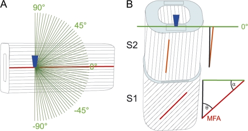
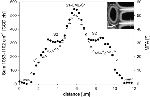
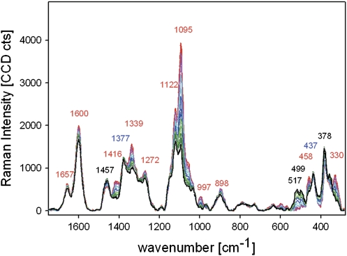
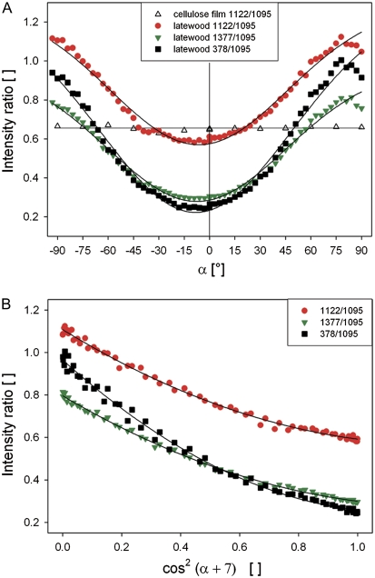
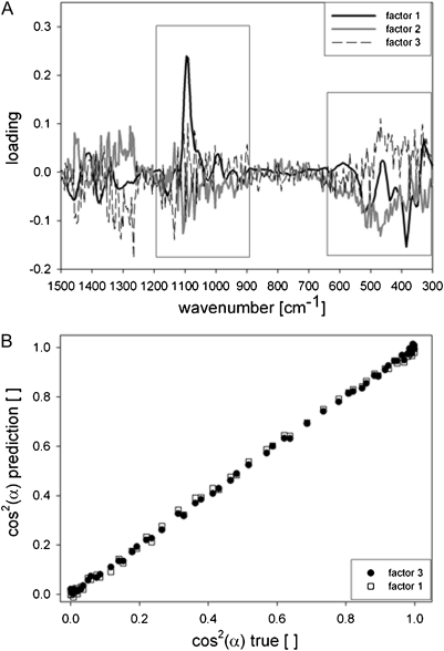
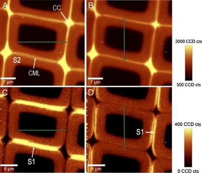
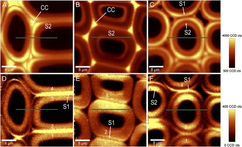
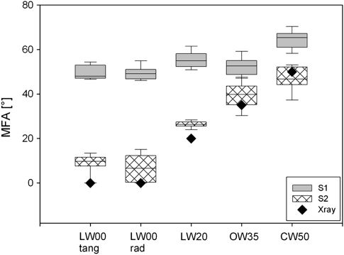
The functional characteristics of plant cell walls depend on the composition of the cell wall polymers, as well as on their highly ordered architecture at scales from a few nanometres to several microns. Raman spectra of wood acquired with linear polarized laser light include information about polymer composition as well as the alignment of cellulose microfibrils with respect to the fibre axis (microfibril angle). By changing the laser polarization direction in 3 degrees steps, the dependency between cellulose and laser orientation direction was investigated. Orientation-dependent changes of band height ratios and spectra were described by quadratic linear regression and partial least square regressions, respectively. Using the models and regressions with high coefficients of determination (R(2) > 0.99) microfibril orientation was predicted in the S1 and S2 layers distinguished by the Raman imaging approach in cross-sections of spruce normal, opposite, and compression wood. The determined microfibril angle (MFA) in the different S2 layers ranged from 0 degrees to 49.9 degrees and was in coincidence with X-ray diffraction determination. With the prerequisite of geometric sample and laser alignment, exact MFA prediction can complete the picture of the chemical cell wall design gained by the Raman imaging approach at the micron level in all plant tissues.

摘要

植物细胞壁的功能特性取决于细胞壁聚合物的组成,以及它们在从几个纳米到几个微米的尺度上的高度有序结构。用线偏振激光光获得的木材拉曼光谱包括关于聚合物组成以及纤维素微纤维相对于纤维轴(微纤维角)排列的信息。通过以 3 度的步长改变激光偏振方向,研究了纤维素和激光取向方向之间的依赖性。通过二次线性回归和偏最小二乘回归分别描述了带高比和光谱的取向依赖性变化。使用具有高决定系数(R(2) > 0.99)的模型和回归,通过在云杉正常、相反和压缩木材的横截面中使用 Raman 成像方法区分的 S1 和 S2 层,预测微纤维取向。在不同的 S2 层中确定的微纤维角(MFA)范围从 0 度到 49.9 度,与 X 射线衍射测定结果一致。在几何样品和激光对准的前提下,准确的 MFA 预测可以完成在所有植物组织中通过 Raman 成像方法在微米级水平上获得的细胞壁化学设计的图像。

https://cdn.ncbi.nlm.nih.gov/pmc/blobs/5576/2803219/8e7e4669f559/jexboterp325f08_ht.jpg
https://cdn.ncbi.nlm.nih.gov/pmc/blobs/5576/2803219/c279324273b2/jexboterp325f01_3c.jpg
https://cdn.ncbi.nlm.nih.gov/pmc/blobs/5576/2803219/71f98abfc7f2/jexboterp325f02_3c.jpg
https://cdn.ncbi.nlm.nih.gov/pmc/blobs/5576/2803219/217c9de4346b/jexboterp325f03_3c.jpg
https://cdn.ncbi.nlm.nih.gov/pmc/blobs/5576/2803219/5b7cc6e10641/jexboterp325f04_ht.jpg
https://cdn.ncbi.nlm.nih.gov/pmc/blobs/5576/2803219/109ddd533a36/jexboterp325f05_3c.jpg
https://cdn.ncbi.nlm.nih.gov/pmc/blobs/5576/2803219/7e02f5350bd5/jexboterp325f06_3c.jpg
https://cdn.ncbi.nlm.nih.gov/pmc/blobs/5576/2803219/52eea0d15b3e/jexboterp325f07_ht.jpg
https://cdn.ncbi.nlm.nih.gov/pmc/blobs/5576/2803219/8e7e4669f559/jexboterp325f08_ht.jpg

相似文献

1
Cellulose microfibril orientation of Picea abies and its variability at the micron-level determined by Raman imaging.由拉曼成像确定的云杉纤维素微纤丝取向及其在微米级的可变性。
J Exp Bot. 2010;61(2):587-95. doi: 10.1093/jxb/erp325. Epub 2009 Dec 9.
2
Non-invasive imaging of cellulose microfibril orientation within plant cell walls by polarized Raman microspectroscopy.利用偏振拉曼显微光谱对植物细胞壁内纤维素微纤丝取向进行非侵入性成像。
Biotechnol Bioeng. 2016 Jan;113(1):82-90. doi: 10.1002/bit.25690. Epub 2015 Sep 18.
3
Role of microfibril angle in molecular deformation of cellulose fibrils in Pinus massoniana compression wood and opposite wood studied by in-situ WAXS.原位 WAXS 研究马尾松压缩木和对应木纤维素纤维分子变形中微纤丝角的作用。
Carbohydr Polym. 2024 Jun 15;334:122024. doi: 10.1016/j.carbpol.2024.122024. Epub 2024 Mar 6.
4
Cellulose microfibril angle in the cell wall of wood fibres.木纤维细胞壁中的纤维素微纤丝角
Biol Rev Camb Philos Soc. 2004 May;79(2):461-72. doi: 10.1017/s1464793103006377.
5
Radial microfibril arrangements in wood cell walls.木细胞壁中径向微纤丝的排列。
Planta. 2022 Sep 10;256(4):75. doi: 10.1007/s00425-022-03976-2.
6
Moisture changes in the plant cell wall force cellulose crystallites to deform.植物细胞壁中的水分变化迫使纤维素微晶发生变形。
J Struct Biol. 2010 Aug;171(2):133-41. doi: 10.1016/j.jsb.2010.04.013. Epub 2010 May 8.
7
Structural variation of tracheids in Norway spruce (Picea abies [L.] Karst.).挪威云杉(Picea abies [L.] Karst.)管胞的结构变异
J Struct Biol. 2001 Nov;136(2):101-9. doi: 10.1006/jsbi.2001.4434.
8
Functional plant cell wall design revealed by the Raman imaging approach.拉曼成像方法揭示的功能性植物细胞壁设计。
Planta. 2011 Apr;233(4):763-72. doi: 10.1007/s00425-010-1338-z. Epub 2011 Jan 1.
9
Cellulose structure and lignin distribution in normal and compression wood of the Maidenhair tree (Ginkgo biloba L.).银杏正常材和压缩材的纤维素结构和木质素分布。
J Integr Plant Biol. 2015 Apr;57(4):388-95. doi: 10.1111/jipb.12349.
10
Raman imaging to investigate ultrastructure and composition of plant cell walls: distribution of lignin and cellulose in black spruce wood (Picea mariana).拉曼成像技术用于研究植物细胞壁的超微结构和组成:黑云杉木(白云杉)中木质素和纤维素的分布
Planta. 2006 Oct;224(5):1141-53. doi: 10.1007/s00425-006-0295-z. Epub 2006 Jun 8.

引用本文的文献

1
Chemical Fingerprint Imaging with Broadband Coherent Anti-Stokes Raman Scattering Microscopy.宽带相干反斯托克斯拉曼散射显微镜的化学指纹成像
Anal Chem. 2025 Aug 12;97(31):16868-16876. doi: 10.1021/acs.analchem.5c01980. Epub 2025 Jul 28.
2
Large Anisotropy of Thermal Conductivity in Oriented Cellulose-Clay Composites.取向纤维素-粘土复合材料中热导率的大各向异性
ACS Omega. 2025 Jun 16;10(25):26560-26566. doi: 10.1021/acsomega.5c00316. eCollection 2025 Jul 1.
3
Valorization of Eichhornia crassipes for the production of cellulose nanocrystals further investigation of plethoric biobased resource.

本文引用的文献

1
Actual versus apparent within cell wall variability of nanoindentation results from wood cell walls related to cellulose microfibril angle.与纤维素微纤丝角相关的木材细胞壁纳米压痕结果在细胞壁内的实际变异性与表观变异性对比
J Mater Sci. 2009;44(16):4399-4406. doi: 10.1007/s10853-009-3665-7. Epub 2009 Aug 1.
2
Arabidopsis cortical microtubules position cellulose synthase delivery to the plasma membrane and interact with cellulose synthase trafficking compartments.拟南芥皮层微管将纤维素合酶传递至质膜并与纤维素合酶运输区室相互作用。
Nat Cell Biol. 2009 Jul;11(7):797-806. doi: 10.1038/ncb1886. Epub 2009 Jun 14.
3
Raman microprobe evidence for lignin orientation in the cell walls of native woody tissue.
凤眼蓝的增值利用生产纤维素纳米晶体进一步研究丰富的生物基资源。
Sci Rep. 2024 May 29;14(1):12387. doi: 10.1038/s41598-024-62159-z.
4
Hydrolyzable tannins are incorporated into the endocarp during sclerification of the water caltrop Trapa natans.可水解单宁在水菱角 Trapa natans 的硬化过程中被纳入内果皮。
Plant Physiol. 2023 Dec 30;194(1):94-105. doi: 10.1093/plphys/kiad408.
5
Spatial consistency of cell growth direction during organ morphogenesis requires CELLULOSE SYNTHASE INTERACTIVE1.器官形态发生过程中细胞生长方向的空间一致性需要纤维素合成酶相互作用蛋白 1。
Cell Rep. 2023 Jul 25;42(7):112689. doi: 10.1016/j.celrep.2023.112689. Epub 2023 Jun 22.
6
Electron tomography unravels new insights into fiber cell wall nanostructure; exploring 3D macromolecular biopolymeric nano-architecture of spruce fiber secondary walls.电子断层扫描揭示了纤维细胞壁纳米结构的新见解;探索云杉纤维次生壁的 3D 大分子生物聚合物纳米结构。
Sci Rep. 2023 Feb 9;13(1):2350. doi: 10.1038/s41598-023-29113-x.
7
Intra-annual fluctuation in morphology and microfibril angle of tracheids revealed by novel microscopy-based imaging.基于新型显微镜成像技术揭示的导管形态和微纤丝角的年际波动。
PLoS One. 2022 Nov 15;17(11):e0277616. doi: 10.1371/journal.pone.0277616. eCollection 2022.
8
The cutin polymer matrix undergoes a fine architectural tuning from early tomato fruit development to ripening.角质聚合物基质在番茄果实早期发育到成熟过程中经历精细的结构调整。
Plant Physiol. 2022 Oct 27;190(3):1821-1840. doi: 10.1093/plphys/kiac392.
9
Visualising lignin quantitatively in plant cell walls by micro-Raman spectroscopy.利用显微拉曼光谱对植物细胞壁中的木质素进行定量可视化分析。
RSC Adv. 2021 Apr 7;11(22):13124-13129. doi: 10.1039/d1ra01825f.
10
A Guide to Elucidate the Hidden Multicomponent Layered Structure of Plant Cuticles by Raman Imaging.拉曼成像解析植物角质层隐藏的多组分层状结构指南
Front Plant Sci. 2021 Dec 17;12:793330. doi: 10.3389/fpls.2021.793330. eCollection 2021.
拉曼微探针证据表明木质素在天然木质组织细胞壁中的取向。
Science. 1985 Feb 8;227(4687):636-8. doi: 10.1126/science.227.4687.636.
4
Microtubules and cellulose microfibrils: how intimate is their relationship?微管与纤维素微纤丝:它们的关系有多紧密?
Trends Plant Sci. 2007 Jul;12(7):279-81. doi: 10.1016/j.tplants.2007.06.002. Epub 2007 Jun 25.
5
Tensile and compressive stresses in tracheids are induced by swelling based on geometrical constraints of the wood cell.基于木材细胞的几何限制,管胞中的拉伸应力和压缩应力是由膨胀引起的。
Planta. 2007 Sep;226(4):981-7. doi: 10.1007/s00425-007-0544-9. Epub 2007 Jun 7.
6
Molecular changes during tensile deformation of single wood fibers followed by Raman microscopy.通过拉曼显微镜观察单根木纤维拉伸变形过程中的分子变化。
Biomacromolecules. 2006 Jul;7(7):2077-81. doi: 10.1021/bm060236g.
7
Determination of mechanical strength properties of hemp fibers using near-infrared fourier transform Raman microspectroscopy.使用近红外傅里叶变换拉曼光谱法测定大麻纤维的机械强度特性
Appl Spectrosc. 2006 Jun;60(6):682-91. doi: 10.1366/000370206777670602.
8
Raman imaging to investigate ultrastructure and composition of plant cell walls: distribution of lignin and cellulose in black spruce wood (Picea mariana).拉曼成像技术用于研究植物细胞壁的超微结构和组成:黑云杉木(白云杉)中木质素和纤维素的分布
Planta. 2006 Oct;224(5):1141-53. doi: 10.1007/s00425-006-0295-z. Epub 2006 Jun 8.
9
Chemical imaging of poplar wood cell walls by confocal Raman microscopy.利用共聚焦拉曼显微镜对杨树木细胞壁进行化学成像。
Plant Physiol. 2006 Apr;140(4):1246-54. doi: 10.1104/pp.105.066993. Epub 2006 Feb 17.
10
Cellulose: fascinating biopolymer and sustainable raw material.纤维素:迷人的生物聚合物和可持续原材料。
Angew Chem Int Ed Engl. 2005 May 30;44(22):3358-93. doi: 10.1002/anie.200460587.