Schepens Eye Research Institute and Department of Ophthalmology, Harvard Medical School, 20 Staniford Street Boston, MA 02114, USA.
Exp Eye Res. 2010 Mar;90(3):444-51. doi: 10.1016/j.exer.2009.12.009. Epub 2009 Dec 29.
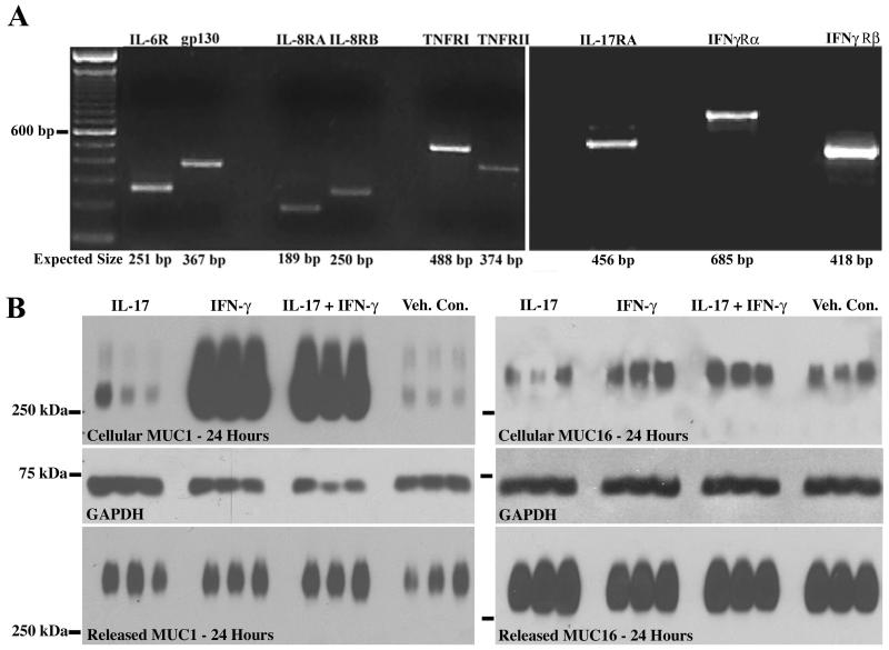
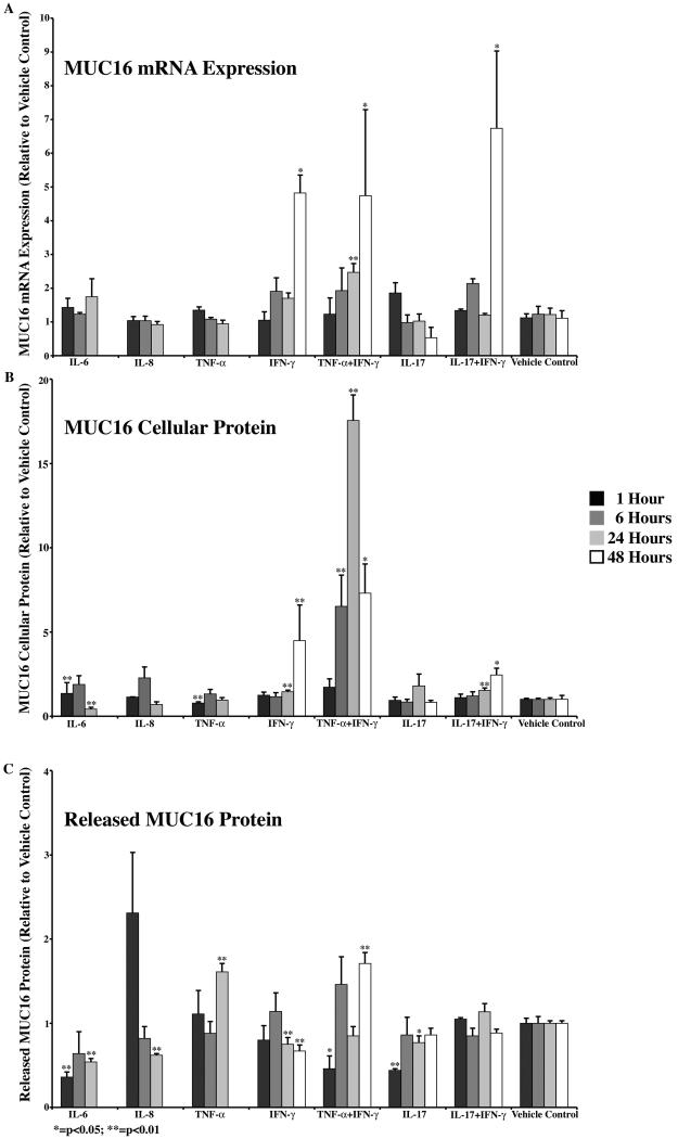
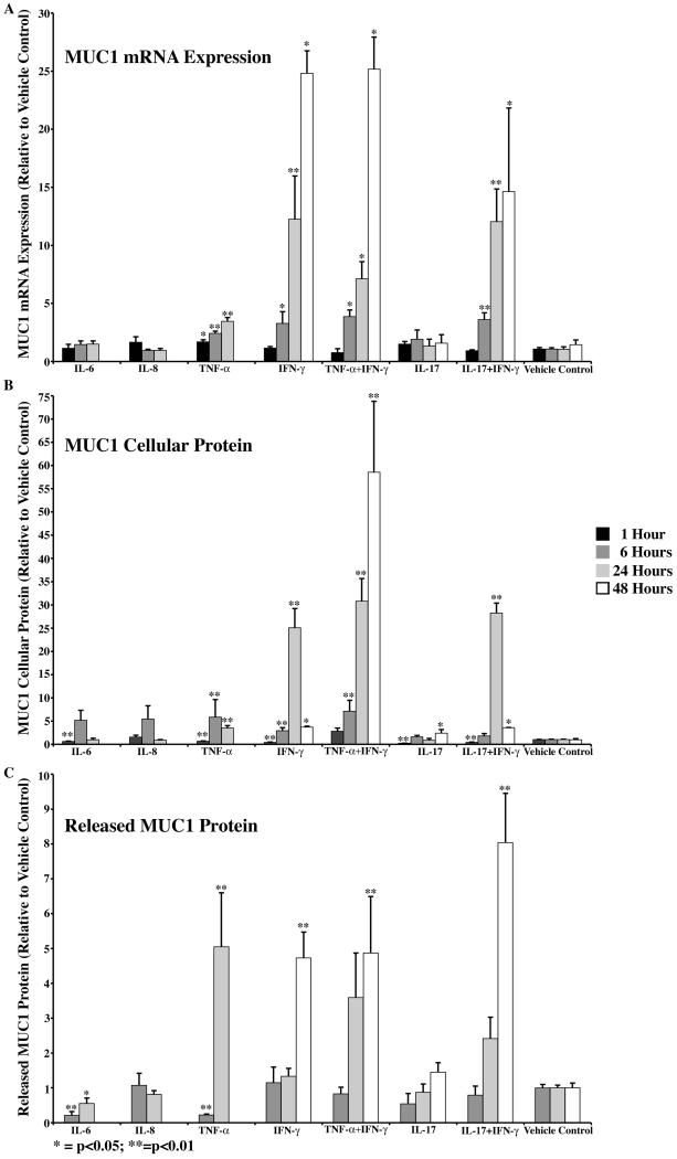
Membrane-associated mucins are altered on the ocular surface in non-Sjögren's dry eye. This study sought to determine if inflammatory mediators, present in tears of dry eye patients, regulate membrane-associated mucins MUC1 and -16 at the level of gene expression, protein biosynthesis and/or ectodomain release. A human corneal limbal epithelial cell line (HCLE), which produces membrane-associated mucins, was used. Cells were treated with interleukin (IL)-6, -8, or -17, tumor necrosis factor-alpha (TNF-alpha), and Interferon-gamma (IFN-gamma), or a combination of TNF-alpha and IFN-gamma, or IFN-gamma and IL-17, for 1, 6, 24, or 48 h. Presence of receptors for these mediators was verified by RT-PCR. Effects of the cytokines on expression levels of MUC1 and -16 were determined by real-time PCR, and on mucin protein biosynthesis and ectodomain release in cell lysates and culture media, respectively, by immunoblot analysis. TNF-alpha and IFN-gamma each significantly induced MUC1 expression, cellular protein content and ectodomain release over time. Combined treatment with the two cytokines was not additive. By comparison, one of the inflammatory mediators, IFN-gamma, affected all three parameters-gene expression, cellular protein, and ectodomain release-for MUC16. Combined treatment with TNF-alpha and IFN-gamma showed effects similar to IFN-gamma alone, except that ectodomain release followed that of TNF-alpha, which induced MUC16 ectodomain release. In conclusion, inflammatory mediators present in tears of dry eye patients can affect MUC1 and -16 on corneal epithelial cells and may be responsible for alterations of surface mucins in dry eye.
眼表非干燥综合征患者的黏蛋白与膜结合发生改变。本研究旨在确定干燥性眼病患者泪液中的炎症介质是否会在基因表达、蛋白生物合成和/或细胞外结构域释放水平上调节膜结合黏蛋白 MUC1 和 -16。我们使用产生膜结合黏蛋白的人角膜缘上皮细胞系(HCLE)。用白细胞介素(IL)-6、-8 或 -17、肿瘤坏死因子-α(TNF-α)和干扰素-γ(IFN-γ),或 TNF-α和 IFN-γ,或 IFN-γ和 IL-17 处理细胞 1、6、24 或 48 小时。通过 RT-PCR 验证这些介质的受体存在。通过实时 PCR 确定细胞因子对 MUC1 和 -16 表达水平的影响,通过免疫印迹分析分别确定对细胞裂解物和培养基中黏蛋白蛋白生物合成和细胞外结构域释放的影响。TNF-α和 IFN-γ均随时间推移显著诱导 MUC1 表达、细胞内蛋白含量和细胞外结构域释放。两种细胞因子的联合治疗没有叠加作用。相比之下,炎症介质之一 IFN-γ影响 MUC16 的所有三个参数-基因表达、细胞内蛋白和细胞外结构域释放。TNF-α和 IFN-γ联合治疗的效果与 IFN-γ单独治疗相似,只是细胞外结构域释放遵循诱导 MUC16 细胞外结构域释放的 TNF-α。总之,干燥性眼病患者泪液中的炎症介质可影响角膜上皮细胞上的 MUC1 和 -16,并可能导致干燥性眼病表面黏蛋白的改变。