Experimental Cardiovascular Medicine, Bristol Heart Institute, University of Bristol, Upper Maudlin Street, Bristol, United Kingdom.
Circ Res. 2010 Mar 5;106(4):757-68. doi: 10.1161/CIRCRESAHA.109.207449. Epub 2010 Jan 7.
Phosphoinositide 3-kinase (PI3K)gamma is expressed in hematopoietic cells, endothelial cells (ECs), and cardiomyocytes and regulates different cellular functions relevant to inflammation, tissue remodeling and cicatrization. Recently, PI3Kgamma inhibitors have been indicated for the treatment of chronic inflammatory/autoimmune diseases and atherosclerosis.
We aimed to determine PI3Kgamma contribution to the angiogenic capacity of ECs and the effect of PI3Kgamma inhibition on healing of myocardial infarction (MI).
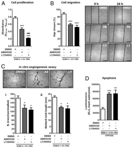
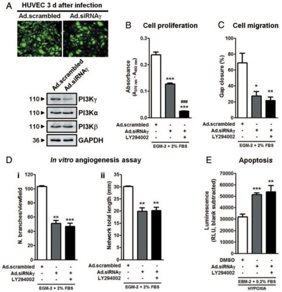
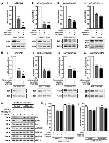
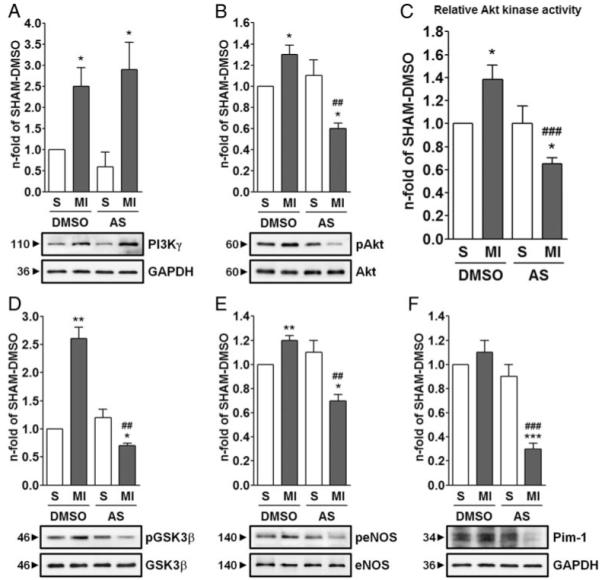
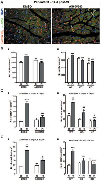
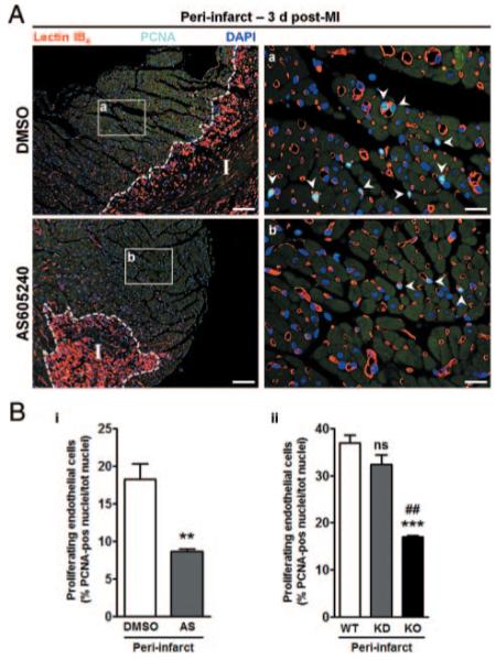
Human umbilical ECs were treated with a selective PI3Kgamma inhibitor, AS605240, or a pan-phosphoinositide 3-kinases inhibitor, LY294002. Both inhibitory treatments and small interfering RNA-mediated PI3Kgamma knockdown strongly impaired ECs angiogenic capacity, because of suppression of the PI3K/Akt and mitogen-activated protein kinase pathways. Constitutive activation of Akt rescued the angiogenic defect. Reparative angiogenesis was studied in vivo in a model of MI. AS605240 did not affect MI-induced PI3Kgamma upregulation, whereas it suppressed Akt activation and downstream signaling. AS605240 strongly reduced inflammation, enhanced cardiomyocyte apoptosis, and impaired survival and proliferation of ECs in peri-infarct zone, which resulted in defective reparative neovascularization. As a consequence, AS605240-treated MI hearts showed increased infarct size and impaired recovery of left ventricular function. Similarly, PI3Kgamma-deficient mice showed impaired reparative neovascularization, enhanced cardiomyocyte apoptosis and marked deterioration of cardiac function following MI. Mice expressing catalytically inactive PI3Kgamma also failed to mount a proper neovascularization, although cardiac dysfunction was similar to wild-type controls.
PI3Kgamma expression and catalytic activity are involved at different levels in reparative neovascularization and healing of MI.
磷酸肌醇 3-激酶 (PI3K)γ在造血细胞、内皮细胞 (EC) 和心肌细胞中表达,并调节与炎症、组织重塑和瘢痕形成相关的不同细胞功能。最近,PI3Kγ抑制剂已被用于治疗慢性炎症/自身免疫性疾病和动脉粥样硬化。
我们旨在确定 PI3Kγ对 EC 血管生成能力的贡献以及 PI3Kγ 抑制对心肌梗死 (MI) 愈合的影响。
用人选择性 PI3Kγ抑制剂 AS605240 或泛磷酸肌醇 3-激酶抑制剂 LY294002 处理人脐静脉 EC。两种抑制性处理和小干扰 RNA 介导的 PI3Kγ 敲低均强烈抑制 EC 的血管生成能力,因为抑制了 PI3K/Akt 和丝裂原活化蛋白激酶途径。Akt 的组成性激活挽救了血管生成缺陷。在 MI 的模型中研究了体内修复性血管生成。AS605240 不影响 MI 诱导的 PI3Kγ上调,但其抑制 Akt 激活和下游信号。AS605240 强烈减少炎症,增强心肌细胞凋亡,并损害梗死周围区 EC 的存活和增殖,导致修复性新生血管形成缺陷。因此,AS605240 处理的 MI 心脏显示出更大的梗死面积和左心室功能恢复受损。同样,PI3Kγ 缺陷型小鼠在 MI 后表现出修复性新生血管形成受损、心肌细胞凋亡增加和心脏功能明显恶化。表达无催化活性的 PI3Kγ的小鼠也未能形成适当的新生血管,尽管心脏功能障碍与野生型对照相似。
PI3Kγ 的表达和催化活性在修复性新生血管形成和 MI 愈合中处于不同的水平。