Université de Lyon, Université Claude Bernard Lyon 1, UPSP 2007,03,135 RTI2B, Lyon, France.
BMC Med Imaging. 2010 Jan 20;10:3. doi: 10.1186/1471-2342-10-3.
The purposes of the study were to determine the relevance and validity of in vivo non-invasive radiographic assessment of the CCLT (Cranial Cruciate Ligament Transection) rabbit model of osteoarthritis (OA) and to estimate the pertinence, reliability and reproducibility of a radiographic OA (ROA) grading scale and associated radiographic atlas.
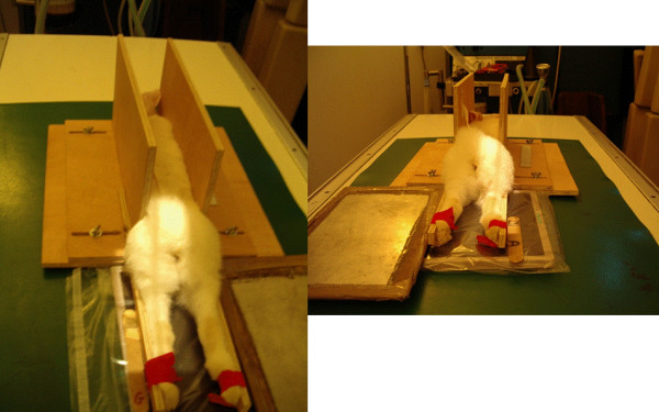
In vivo non-invasive extended non weight-bearing radiography of the rabbit femorotibial joint was standardized. Two hundred and fifty radiographs from control and CCLT rabbits up to five months after surgery were reviewed by three readers. They subsequently constructed an original semi-quantitative grading scale as well as an illustrative atlas of individual ROA feature for the medial compartment. To measure agreements, five readers independently scored the same radiographic sample using this atlas and three of them performed a second reading. To evaluate the pertinence of the ROA grading scale, ROA results were compared with gross examination in forty operated and ten control rabbits.
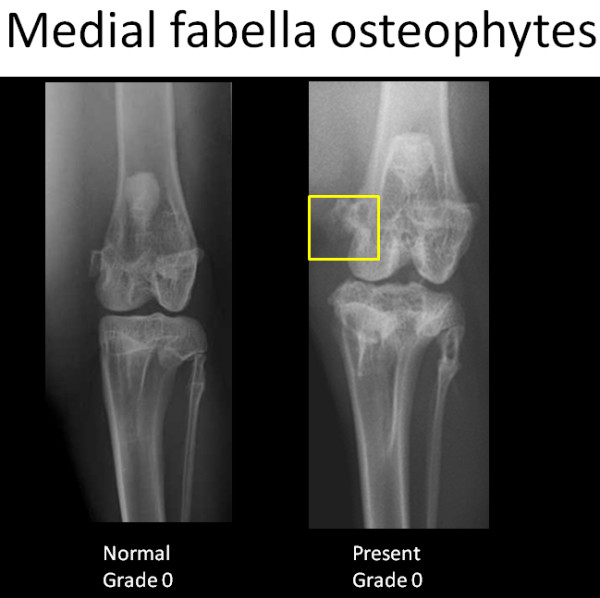
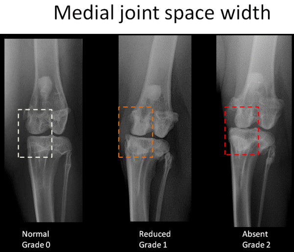
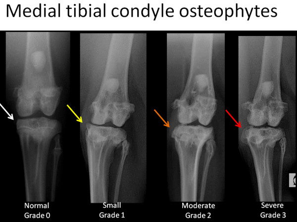
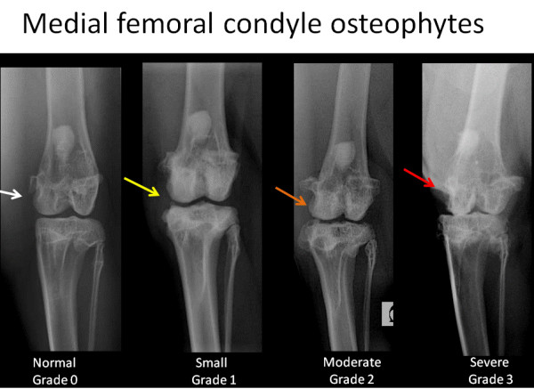
Radiographic osteophytes of medial femoral condyles and medial tibial condyles were scored on a four point scale and dichotomously for osteophytes of medial fabella. Medial joint space width was scored as normal, reduced or absent. Each ROA features was well correlated with gross examination (p < 0.001). ICCs of each ROA features demonstrated excellent agreement between readers and within reading. Global ROA score gave the highest ICCs value for between (ICC 0.93; CI 0.90-0.96) and within (ICC ranged from 0.94 to 0.96) observer agreements. Among all individual ROA features, medial joint space width scoring gave the highest overall reliability and reproducibility and was correlated with both meniscal and cartilage macroscopic lesions (rs = 0.68 and rs = 0.58, p < 0.001 respectively). Radiographic osteophytes of the medial femoral condyle gave the lowest agreements while being well correlated with the macroscopic osteophytes (rs = 0.64, p < 0.001).
Non-invasive in vivo radiography of the rabbit femorotibial joint is feasible, relevant and allows a reproducible grading of experimentally induced OA lesion. The radiographic grading scale and atlas presented could be used as a template for in vivo non invasive grading of ROA in preclinical studies and could allow future comparisons between studies.
本研究旨在确定 CCLT(十字韧带切断)兔骨关节炎(OA)模型的体内非侵入性放射学评估的相关性和有效性,并评估放射学 OA(ROA)分级量表及其相关放射学图谱的相关性、可靠性和可重复性。
对兔股胫关节进行标准化的非侵入性、非负重的扩展放射摄影。对手术后 5 个月内的 250 张对照和 CCLT 兔的 X 线片进行了 3 位读者的回顾性分析。随后,他们构建了一个原始的半定量分级量表和内侧间室的个体 ROA 特征的说明性图谱。为了评估一致性,5 位读者使用该图谱独立地对同一批 X 线片进行评分,其中 3 位读者进行了第二次阅读。为了评估 ROA 分级量表的相关性,将 ROA 结果与 40 只手术兔和 10 只对照兔的大体检查结果进行了比较。
对内侧股骨髁和内侧胫骨髁的放射状骨赘进行了四分制评分,并对内侧副髌骨的骨赘进行了二分制评分。内侧关节间隙宽度被评为正常、减少或缺失。每个 ROA 特征均与大体检查高度相关(p<0.001)。每个 ROA 特征的 ICC 均显示读者之间和阅读内具有极好的一致性。全局 ROA 评分在观察者之间(ICC 0.93;CI 0.90-0.96)和观察者内(ICC 范围为 0.94 至 0.96)均给出了最高的 ICC 值。在所有的 ROA 特征中,内侧关节间隙宽度评分具有最高的总体可靠性和可重复性,与半月板和软骨的大体病变相关(rs=0.68 和 rs=0.58,p<0.001)。内侧股骨髁的放射状骨赘的一致性最低,但与大体骨赘高度相关(rs=0.64,p<0.001)。
兔股胫关节的非侵入性活体放射摄影是可行的、相关的,并且可以对实验性诱导的 OA 病变进行可重复的分级。所提出的放射学分级量表和图谱可作为临床前研究中 ROA 活体非侵入性分级的模板,并可允许未来在研究之间进行比较。