Department of Radiology, UMDNJ-New Jersey Medical School, Newark, NJ 07103, USA.
Magn Reson Imaging. 2010 May;28(4):466-76. doi: 10.1016/j.mri.2009.12.007. Epub 2010 Feb 1.
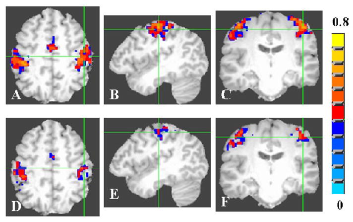
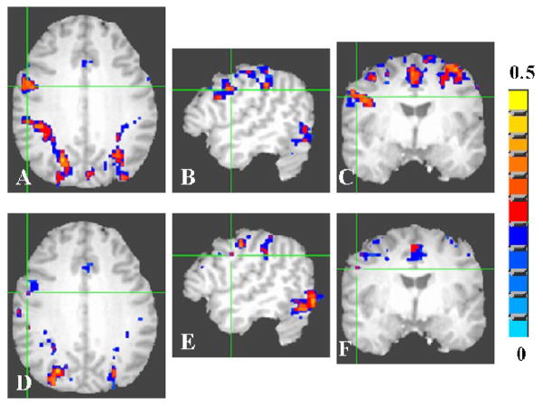
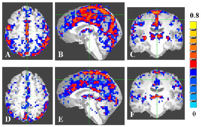
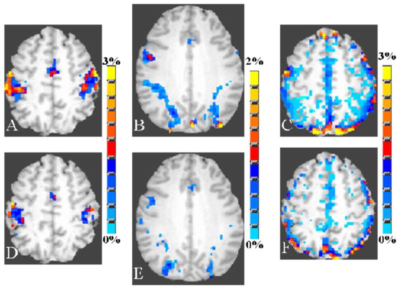
Neural, vascular and structural variables contributing to the blood oxygen level-dependent (BOLD) signal response variability were investigated in younger and older humans. Twelve younger healthy human subjects (six male and six female; mean age: 24 years; range: 19-27 years) and 12 older healthy subjects (five male and seven female; mean age: 58 years; range: 55-71 years) with no history of head trauma and neurological disease were scanned. Functional magnetic resonance imaging measurements using the BOLD contrast were made when participants performed a motor, cognitive or a breath hold (BH) task. Activation volume and the BOLD response amplitude were estimated for the younger and older at both group and subject levels. Mean activation volume was reduced by 45%, 40% and 38% in the elderly group during the motor, cognitive and BH tasks, respectively, compared to the younger. Reduction in activation volume was substantially higher compared to the reduction in the gray matter volume of 14% in the older compared to the younger. A significantly larger variability in the intersubject BOLD signal change occurred during the motor task, compared to the cognitive task. BH-induced BOLD signal change between subjects was significantly less-variable in the motor task-activated areas in the younger compared to older whereas such a difference between age groups was not observed during the cognitive task. Hemodynamic scaling using the BH signal substantially reduced the BOLD signal variability during the motor task compared to the cognitive task. The results indicate that the origin of the BOLD signal variability between subjects was predominantly vascular during the motor task while being principally a consequence of neural variability during the cognitive task. Thus, in addition to gray matter differences, the type of task performed can have different vascular variability weighting that can influence age-related differences in brain functional response.
探讨了年轻和老年人类中导致血氧水平依赖(BOLD)信号响应变异性的神经、血管和结构变量。 12 名年轻健康的人类受试者(6 名男性和 6 名女性;平均年龄:24 岁;范围:19-27 岁)和 12 名年龄较大的健康受试者(5 名男性和 7 名女性;平均年龄:58 岁;范围:55-71 岁),没有头部外伤和神经系统疾病的病史。当参与者执行运动、认知或呼吸保持(BH)任务时,使用 BOLD 对比进行功能磁共振成像测量。在组和个体水平上,估计了年轻和老年的激活体积和 BOLD 响应幅度。与年轻组相比,在运动、认知和 BH 任务中,老年组的平均激活体积分别减少了 45%、40%和 38%。与年轻组相比,老年组的灰质体积减少了 14%,而激活体积的减少要高得多。在运动任务中,个体间 BOLD 信号变化的变异性明显大于认知任务。与认知任务相比,在运动任务激活区域,个体间 BH 诱导的 BOLD 信号变化的变异性明显较小。使用 BH 信号进行血液动力学缩放可显著降低运动任务中 BOLD 信号的变异性,而在认知任务中则降低幅度较小。结果表明,在运动任务中,个体间 BOLD 信号变异性的主要来源是血管,而在认知任务中则主要是神经变异性的结果。因此,除了灰质差异之外,执行的任务类型可以具有不同的血管变异性权重,从而影响大脑功能响应的年龄相关差异。