Department of Orthopaedic Surgery, University of Pennsylvania, Philadelphia, PA 19104, USA.
Biomaterials. 2010 May;31(14):4113-20. doi: 10.1016/j.biomaterials.2010.01.098. Epub 2010 Feb 10.
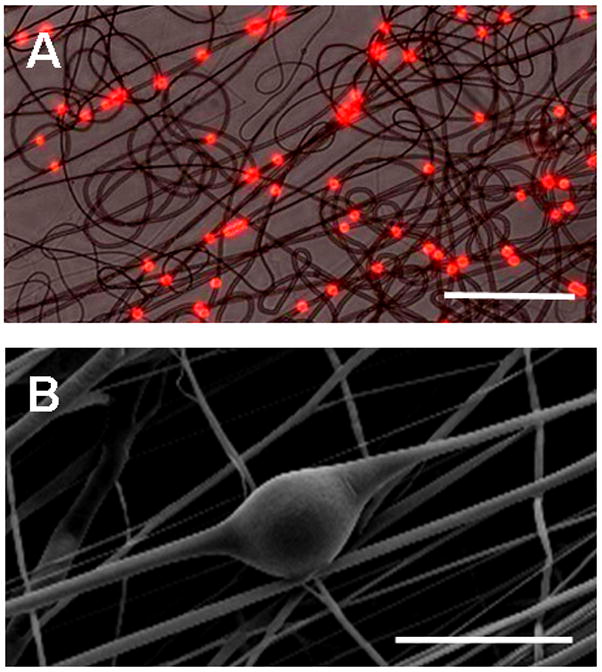
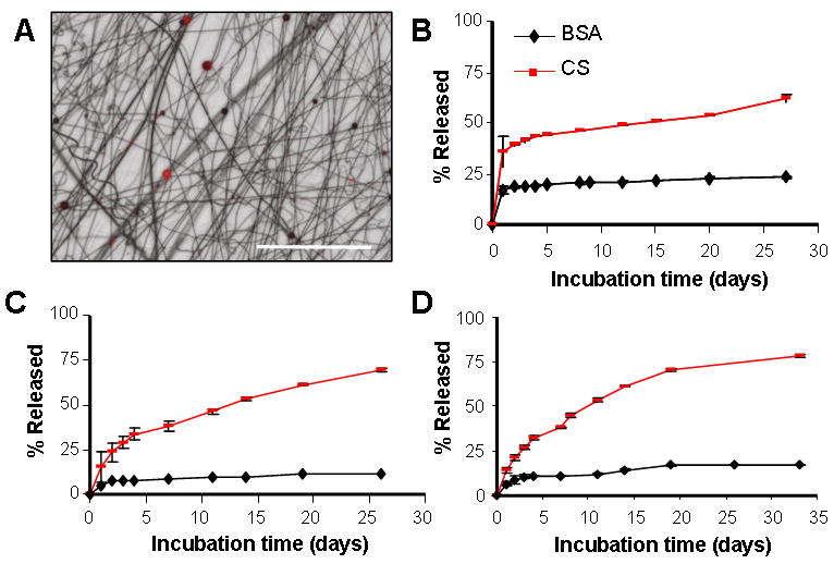
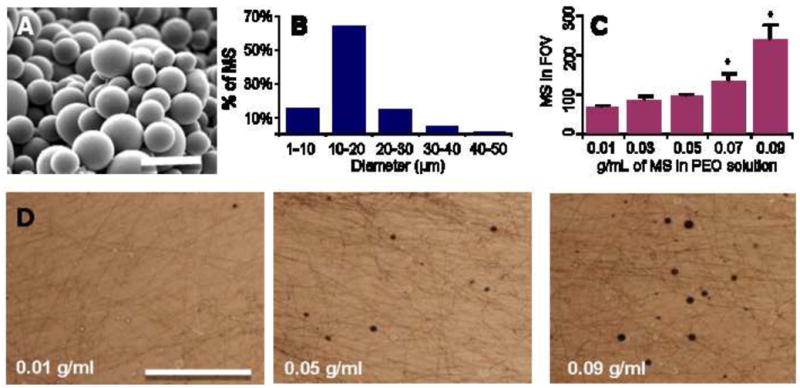
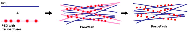
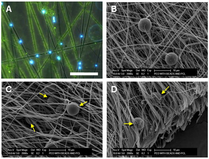
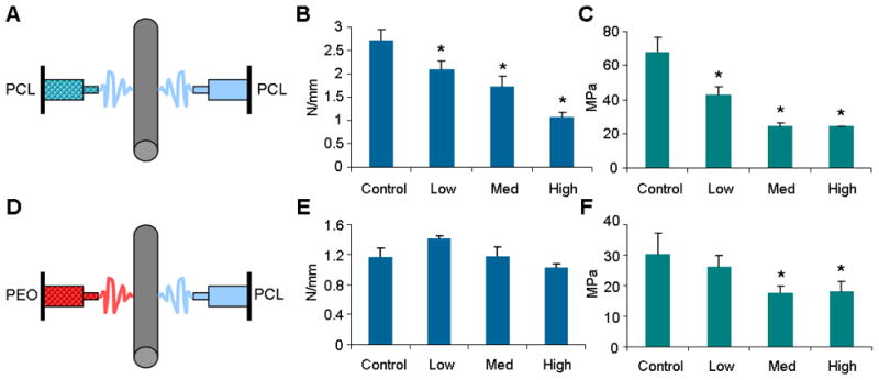
Aligned nanofibrous scaffolds can recapitulate the structural hierarchy of fiber-reinforced tissues of the musculoskeletal system. While these electrospun fibrous scaffolds provide physical cues that can direct tissue formation when seeded with cells, the ability to chemically guide a population of cells, without disrupting scaffold mechanical properties, would improve the maturation of such constructs and add additional functionality to the system both in vitro and in vivo. In this study, we developed a fabrication technique to entrap drug-delivering microspheres within nanofibrous scaffolds. We hypothesized that entrapping microspheres between fibers would have a less adverse impact on mechanical properties than placing microspheres within the fibers themselves, and that the composite would exhibit sustained release of multiple model compounds. Our results show that microspheres ranging from 10 - 20 microns in diameter could be electrospun in a dose-dependent manner to form nanofibrous composites. When delivered in a sacrificial PEO fiber population, microspheres remained securely entrapped between slow-degrading PCL fibers after removal of the sacrificial delivery component. Stiffness and modulus of the composite decreased with increasing microsphere density for composites in which microspheres were entrapped within each fiber, while stiffness did not change when microspheres were entrapped between fibers. The release profiles of the composite structures were similar to free microspheres, with an initial burst release followed by a sustained release of the model molecules over 4 weeks. Further, multiple model molecules were released from a single scaffold composite, demonstrating the capacity for multi-factor controlled release ideal for complex growth factor delivery from these structures.
排列的纳米纤维支架可以再现肌肉骨骼系统纤维增强组织的结构层次。虽然这些静电纺丝纤维支架提供了物理线索,可以在接种细胞时指导组织形成,但能够在不破坏支架机械性能的情况下化学引导细胞群体,将改善这些结构的成熟度,并为体外和体内系统增加额外的功能。在这项研究中,我们开发了一种将载药微球包埋在纳米纤维支架中的制造技术。我们假设将微球包埋在纤维之间对机械性能的不利影响将小于将微球置于纤维内部,并且复合材料将表现出多种模型化合物的持续释放。我们的结果表明,直径为 10-20 微米的微球可以以剂量依赖的方式静电纺丝形成纳米纤维复合材料。当在牺牲的 PEO 纤维群中递送时,微球在去除牺牲的递送组件后仍牢固地包埋在缓慢降解的 PCL 纤维之间。当微球被包埋在每根纤维内时,复合材料的刚度和模量随微球密度的增加而降低,而当微球被包埋在纤维之间时,刚度没有变化。复合结构的释放曲线与游离微球相似,具有初始突释,然后在 4 周内持续释放模型分子。此外,多种模型分子从单个支架复合材料中释放出来,证明了从这些结构中释放多种因子的能力,这是复杂生长因子释放的理想选择。