Department of Chemical and Biomolecular Engineering, Johns Hopkins University, Baltimore, MD 21218, USA.
Cell Death Differ. 2010 Aug;17(8):1325-34. doi: 10.1038/cdd.2010.13. Epub 2010 Feb 12.
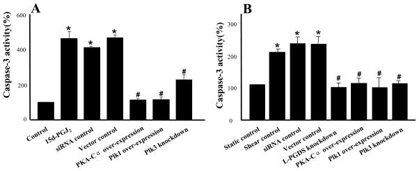
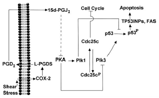
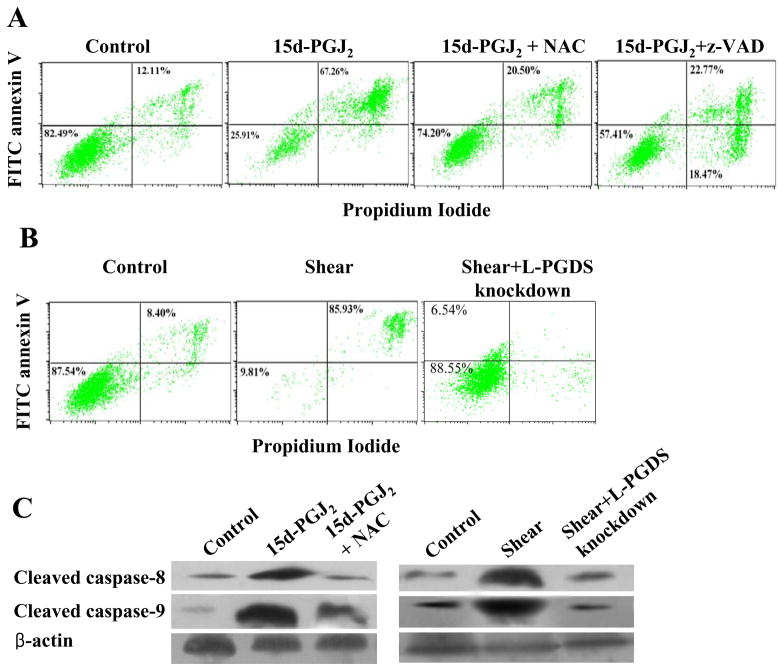
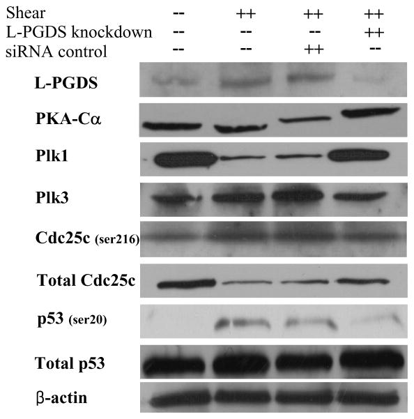
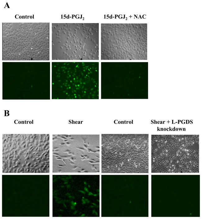
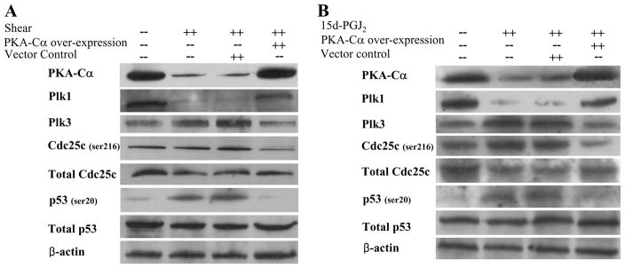
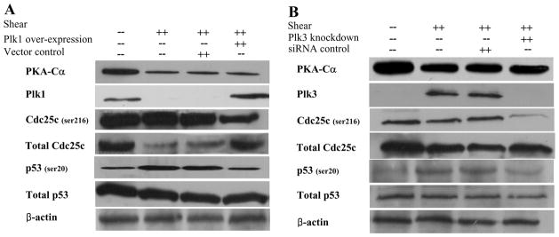
Excessive mechanical loading of cartilage producing hydrostatic stress, tensile strain and fluid flow leads to chondrocyte apoptosis and osteoarthritis. High fluid flow induces cyclooxygenase-2 (COX-2) expression in sheared chondrocytes, which suppresses their antioxidant capacity and contributes to apoptosis. The pivotal role of COX-2 in shear-induced chondrocyte apoptosis and the conflicting literature data on the roles of prostaglandin (PG)E(2), PGD(2) and its metabolite 15-deoxy-Delta(12,14)-PGJ(2) (15d-PGJ(2)) in chondrocyte apoptosis prompted us to analyze which COX-2-derived PG is involved in this process. We show that exogenously added PGD(2) and 15d-PGJ(2), but not PGE(2), diminish the viability of human T/C-28a2 chondrocytes under static conditions. In agreement with these observations, knockdown of L-PGD synthase (L-PGDS) abolishes shear-induced chondrocyte apoptosis. Using cDNA microarrays in conjunction with clustering algorithms, we propose a novel signaling pathway by which high fluid shear mediates COX-2/L-PGDS-dependent chondrocyte apoptosis, which is validated by molecular interventions. We show that L-PGDS controls the downregulation of protein kinase A (PKA), which in turn regulates Polo-like kinase1 (Plk1) and Plk3. Plks target p53, which controls the transcription of p53 effectors (TP53INPs, FAS and Bax) involved in chondrocyte apoptosis. Reconstructing the signaling network regulating chondrocyte apoptosis may provide insights to optimize conditions for culturing artificial cartilage in bioreactors and for developing therapeutic strategies for arthritic disorders.
软骨承受过度的机械负荷会产生静水压力、拉伸应变和流体流动,导致软骨细胞凋亡和骨关节炎。高流体流动会诱导剪切软骨细胞中环氧化酶-2(COX-2)的表达,从而抑制其抗氧化能力并促进细胞凋亡。COX-2 在剪切诱导的软骨细胞凋亡中的关键作用,以及关于前列腺素(PG)E2、PGD2 及其代谢物 15-脱氧-Delta(12,14)-PGJ2(15d-PGJ2)在软骨细胞凋亡中的作用的相互矛盾的文献数据,促使我们分析 COX-2 衍生的 PG 中哪一种参与了这一过程。我们表明,外源性添加 PGD2 和 15d-PGJ2,但不是 PGE2,会降低静息状态下人类 T/C-28a2 软骨细胞的活力。这些观察结果与 L-PGD 合酶(L-PGDS)的敲低消除剪切诱导的软骨细胞凋亡一致。我们使用 cDNA 微阵列结合聚类算法,提出了一种新的信号通路,即高流体剪切通过 COX-2/L-PGDS 依赖性软骨细胞凋亡,通过分子干预进行了验证。我们表明,L-PGDS 控制蛋白激酶 A(PKA)的下调,而 PKA 反过来又调节 Polo 样激酶 1(Plk1)和 Plk3。Plks 靶向 p53,控制参与软骨细胞凋亡的 p53 效应物(TP53INPs、FAS 和 Bax)的转录。重建调控软骨细胞凋亡的信号网络可能为优化生物反应器中人工软骨培养条件和开发关节炎疾病治疗策略提供见解。