Institut Nationale de la Santé et de la Recherche Médicale (INSERM), U846, Stem Cell and Brain Research Institute, Department of Chronobiology, F-69500, Bron, France.
J Comp Neurol. 2010 Apr 15;518(8):1249-63. doi: 10.1002/cne.22272.
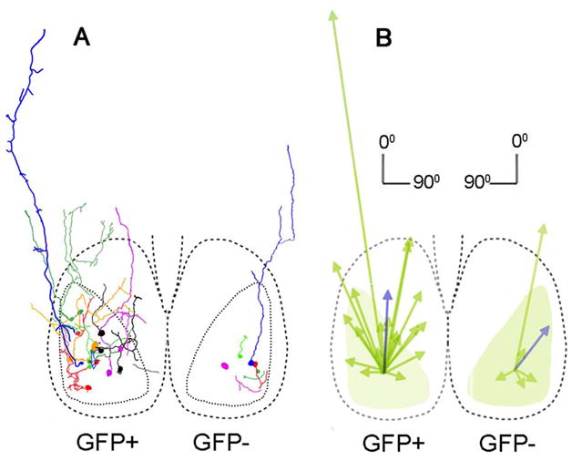
The suprachiasmatic nucleus (SCN) of the hypothalamus regulates daily rhythms in physiology and behavior. It is composed of a heterogeneous population of cells that together form the circuits underlying its master clock function. Numerous studies suggest the existence of two regions that have been termed core and shell. At a gross level, differences between these regions map to distinct functional differences, although the specific role(s) of various peptidergic cellular phenotypes remains unknown. In mouse, gastrin-releasing peptide (GRP) cells lie in the core, are directly retinorecipient, and lack detectable rhythmicity in clock gene expression, raising interest in their role in the SCN. Here, we provide evidence that calbindin-expressing cells of perinatal mouse SCN express GRP, identified by a green fluorescent protein (GFP+), but lack detectable calbindin later in development. To explore the intra-SCN network in which GRP neurons participate, individual GFP+ cells were filled with tracer and their morphological characteristics, processes, and connections, as well as those of their non-GFP-containing immediate neighbors, were compared. The results show that GFP+ neurons form a dense network of local circuits within the core, revealed by appositions on other GFP+ cells and by the presence of dye-coupled cells. Dendrites and axons of GFP+ cells make appositions on arginine vasopressin neurons, whereas non-GFP cells have a less extensive fiber network, largely confined to the region of GFP+ cells. The results point to specialized circuitry within the SCN, presumably supporting synchronization of neural activity and reciprocal communication between core and shell regions.
视交叉上核(SCN)调节生理和行为的日常节律。它由异质细胞组成,共同形成其主时钟功能的回路。许多研究表明存在两个区域,它们被称为核心和壳。在大体水平上,这些区域之间的差异映射到明显的功能差异,尽管各种肽能细胞表型的具体作用仍不清楚。在小鼠中,胃泌素释放肽(GRP)细胞位于核心,直接接受视网膜输入,并且时钟基因表达中缺乏可检测的节律性,这引起了人们对其在 SCN 中作用的兴趣。在这里,我们提供了证据表明,围产期小鼠 SCN 的 calbindin 表达细胞表达 GRP,通过绿色荧光蛋白(GFP+)鉴定,但在发育后期缺乏可检测的 calbindin。为了探索 GRP 神经元参与的 SCN 内网络,用示踪剂填充单个 GFP+细胞,并比较其形态特征、过程和连接,以及其非 GFP 包含的直接相邻细胞的特征。结果表明,GFP+神经元在核心内形成了一个密集的局部回路网络,通过与其他 GFP+细胞的接触和存在偶联的细胞来揭示。GFP+细胞的树突和轴突与精氨酸加压素神经元接触,而非 GFP 细胞的纤维网络较少,主要局限于 GFP+细胞区域。结果表明 SCN 内存在专门的回路,可能支持神经活动的同步和核心与壳区之间的相互通讯。