NYU Cancer Institute and the NYU Gene Therapy Center, NYU School of Medicine, 550 First Avenue, New York, NY 10016, USA.
Mol Cancer. 2010 Feb 12;9:37. doi: 10.1186/1476-4598-9-37.
Sindbis viral vectors are able to efficiently target and kill tumor cells in vivo, as shown using pancreatic and ovarian cancer models. Infection results in apoptosis both in vitro and in vivo. Sindbis vector uptake is mediated by the LAMR, which is upregulated on a number of different tumor types, thus conferring specificity of the vector to a wide range of cancers. In this study we elucidate the mechanism of apoptosis in two tumor cell lines, MOSEC, derived from the ovarian epithelium and Pan02, derived from a pancreatic adenocarcinoma. A comprehensive understanding of the mechanism of apoptosis would facilitate the design of more effective vectors for cancer therapy.
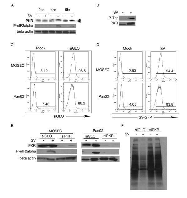
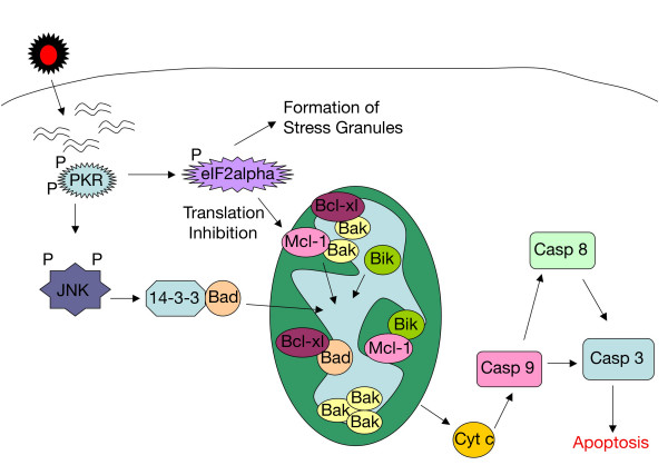
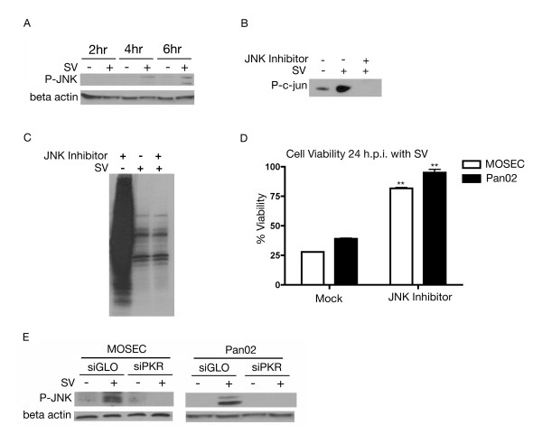
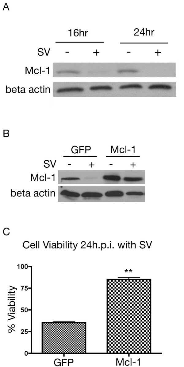
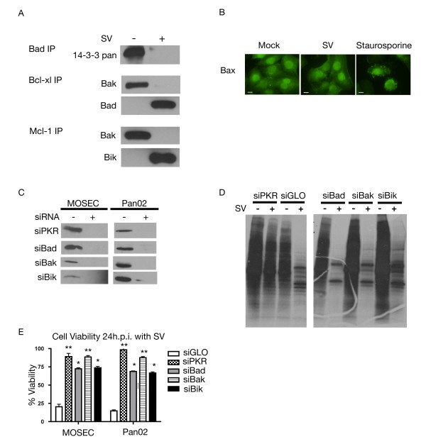
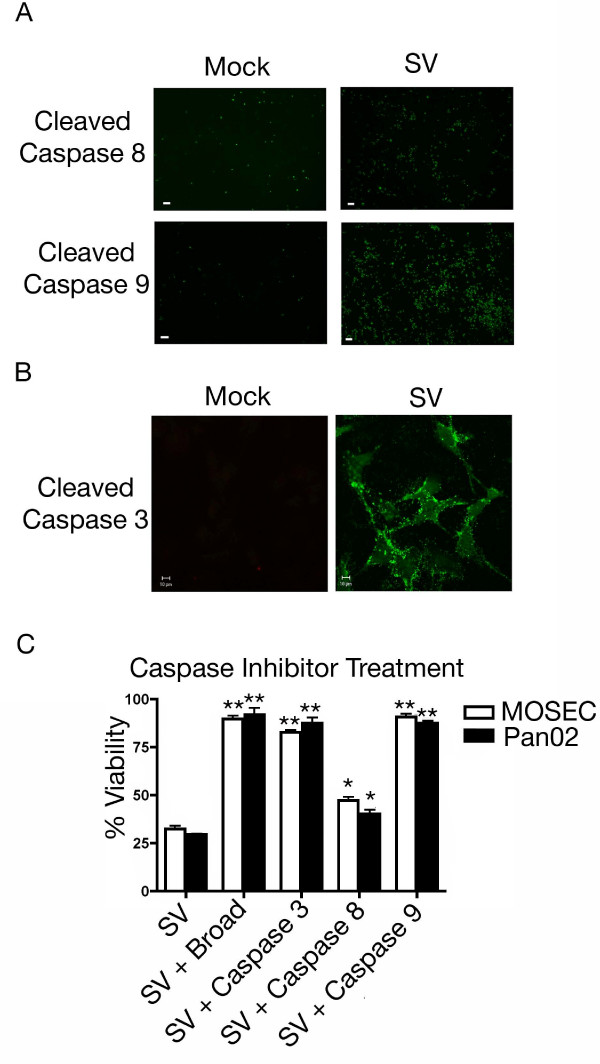
The initial phase of Sindbis vector induced apoptosis in MOSEC and Pan02 models reconfirms that viral infection is sensed by PKR due to double-stranded RNA intermediates associated with genomic replication. PKR activation results in translation inhibition through eIF2alpha phosphorylation and initiation of the stress response. Our studies indicate that the roles of two proteins, Mcl-1 and JNK, intimately link Sindbis induced translational arrest and cellular stress. Translational arrest inhibits the synthesis of anti-apoptotic Bcl-2 protein, Mcl-1. JNK activation triggers the release of Bad from 14-3-3, which ultimately results in apoptosis. These signals from translational arrest and cellular stress are propagated to the mitochondria where Bad and Bik bind to Bcl-xl and Mcl-1 respectively. Formation of these heterodimers displaces Bak, which results in caspase 9 cleavage and signaling through the mitochondrial pathway of apoptosis.
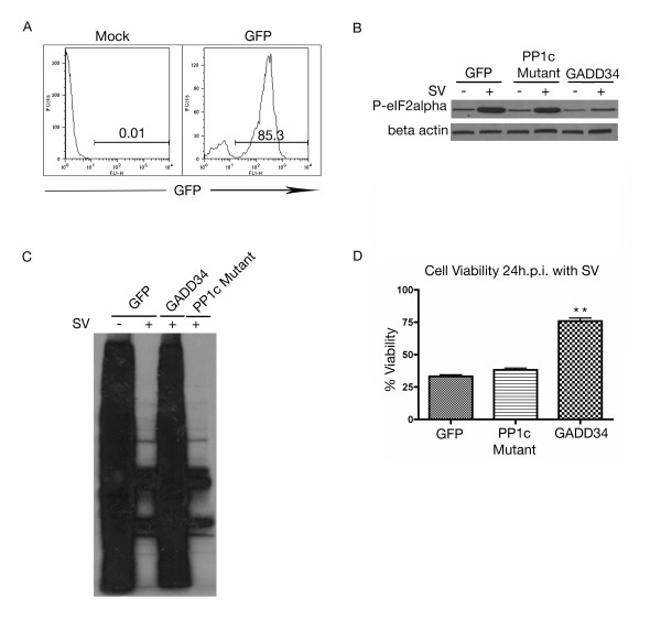
The host cell response to Sindbis is triggered through PKR activation. Our studies demonstrate that PKR activation and subsequent translational arrest is linked to both cellular stress and apoptosis. We have also found the linkage point between translational arrest and apoptosis to be Mcl-1, a protein whose constant translation is required for inhibition of apoptosis. With this information vectors can be designed, which express or repress proteins implicated in this study, to enhance their therapeutic potential.
辛德毕斯病毒载体能够有效地靶向和杀死体内的肿瘤细胞,这已在胰腺和卵巢癌模型中得到证实。感染导致体外和体内的细胞凋亡。辛德毕斯载体的摄取是由 LAMR 介导的,LAMR 在许多不同的肿瘤类型中上调,从而使载体特异性地针对广泛的癌症。在这项研究中,我们阐明了两种肿瘤细胞系(MOSEC,来源于卵巢上皮细胞,和 Pan02,来源于胰腺腺癌)中凋亡的机制。对凋亡机制的全面了解将有助于设计更有效的癌症治疗载体。
辛德毕斯病毒载体诱导 MOSEC 和 Pan02 模型中细胞凋亡的初始阶段再次证实,病毒感染是由于与基因组复制相关的双链 RNA 中间体而被 PKR 感知的。PKR 的激活通过 eIF2alpha 磷酸化导致翻译抑制和应激反应的起始。我们的研究表明,两种蛋白质,Mcl-1 和 JNK,紧密地将辛德毕斯诱导的翻译抑制与细胞应激联系起来。翻译抑制抑制抗凋亡 Bcl-2 蛋白 Mcl-1 的合成。JNK 的激活触发 Bad 从 14-3-3 释放,最终导致细胞凋亡。这些来自翻译抑制和细胞应激的信号传递到线粒体,Bad 和 Bik 分别与 Bcl-xl 和 Mcl-1 结合。这些异二聚体的形成取代了 Bak,导致 caspase 9 的切割和通过线粒体凋亡途径的信号传递。
宿主细胞对辛德毕斯的反应是通过 PKR 的激活触发的。我们的研究表明,PKR 的激活和随后的翻译抑制与细胞应激和凋亡有关。我们还发现,翻译抑制和凋亡之间的连接点是 Mcl-1,这种蛋白质的持续翻译对于抑制凋亡是必需的。有了这些信息,就可以设计出表达或抑制本研究中涉及的蛋白质的载体,以增强它们的治疗潜力。