Cardiovasular Research Institute, Department of Medicine, University of California, San Francisco, San Francisco, CA 94158, USA.
Dev Cell. 2010 Jan 19;18(1):25-38. doi: 10.1016/j.devcel.2009.11.014.
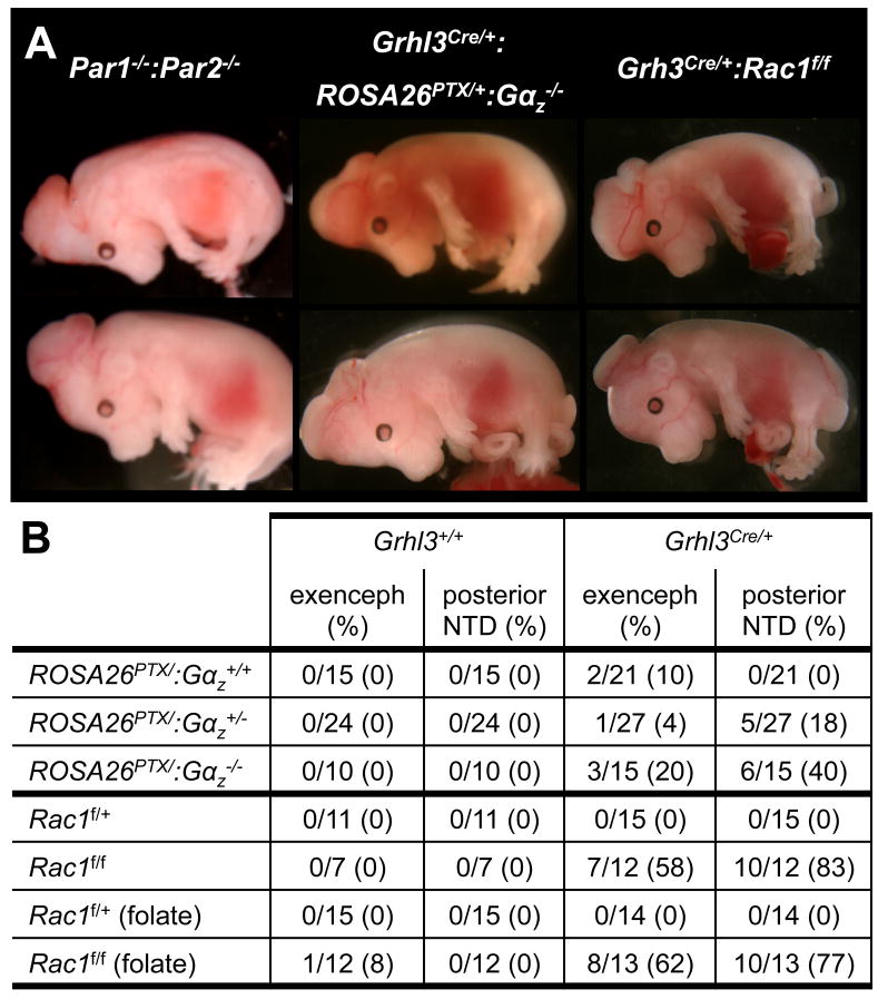
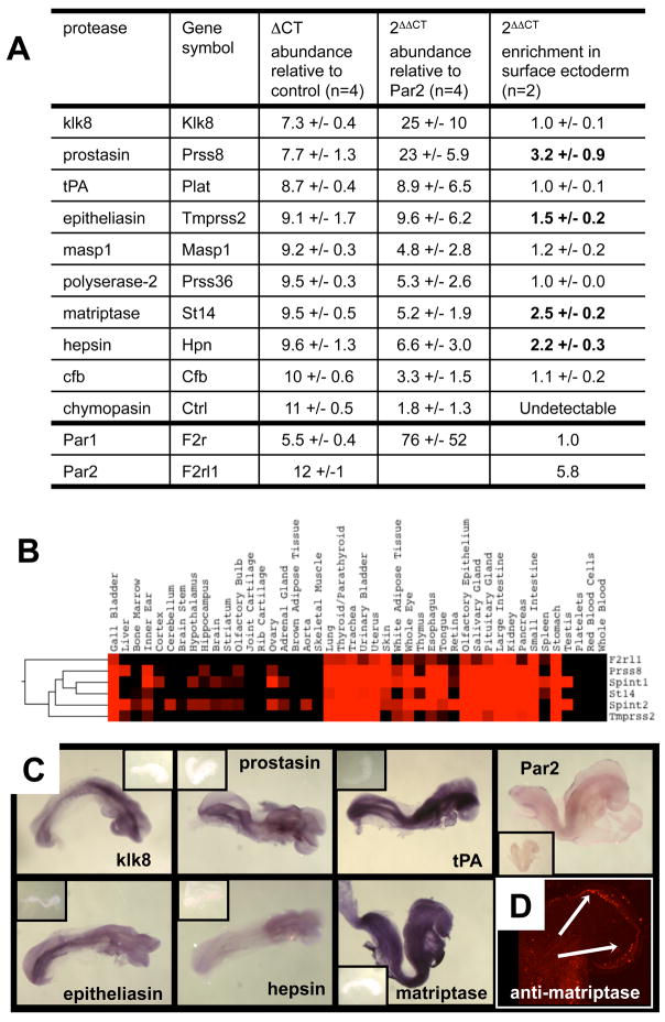
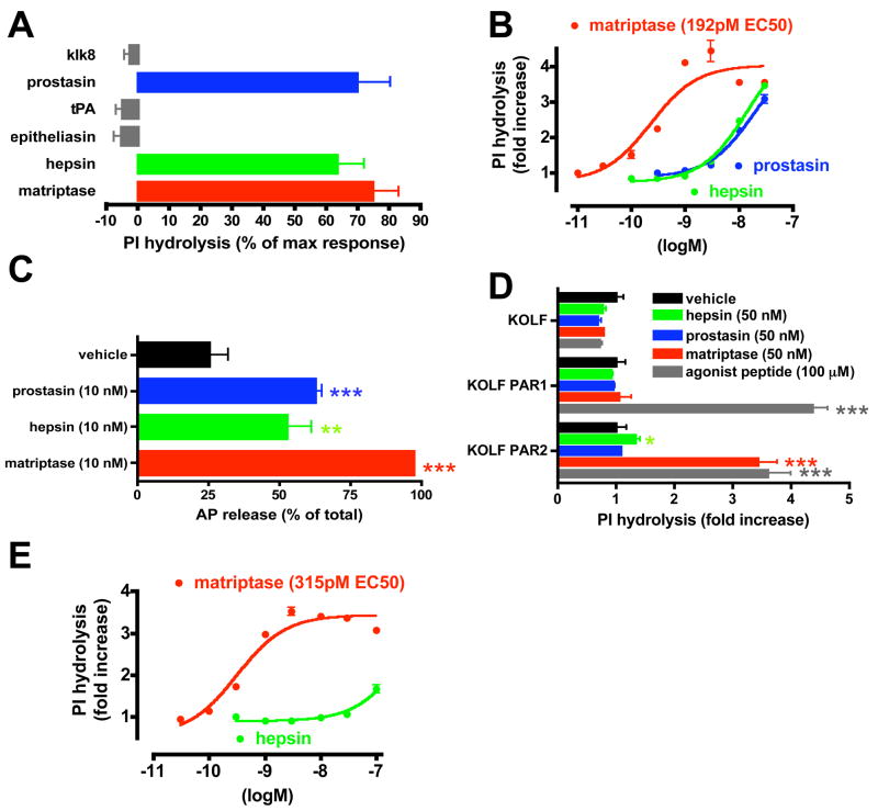
We report an unexpected role for protease signaling in neural tube closure and the formation of the central nervous system. Mouse embryos lacking protease-activated receptors 1 and 2 showed defective hindbrain and posterior neuropore closure and developed exencephaly and spina bifida, important human congenital anomalies. Par1 and Par2 were expressed in surface ectoderm, and Par2 was expressed selectively along the line of closure. Ablation of G(i/z) and Rac1 function in these Par2-expressing cells disrupted neural tube closure, further implicating G protein-coupled receptors and identifying a likely effector pathway. Cluster analysis of protease and Par2 expression patterns revealed a group of membrane-tethered proteases often coexpressed with Par2. Among these, matriptase activated Par2 with picomolar potency, and hepsin and prostasin activated matriptase. Together, our results suggest a role for protease-activated receptor signaling in neural tube closure and identify a local protease network that may trigger Par2 signaling and monitor and regulate epithelial integrity in this context.
我们报道了蛋白酶信号在神经管闭合和中枢神经系统形成中的一个意外作用。缺乏蛋白酶激活受体 1 和 2 的小鼠胚胎表现出后脑和后神经孔闭合缺陷,并发展为无脑畸形和脊柱裂,这是重要的人类先天异常。Par1 和 Par2 在表面外胚层中表达,Par2 沿闭合线选择性表达。这些表达 Par2 的细胞中 G(i/z)和 Rac1 功能的缺失破坏了神经管闭合,进一步表明 G 蛋白偶联受体的作用,并确定了一种可能的效应途径。蛋白酶和 Par2 表达模式的聚类分析显示了一组通常与 Par2 共表达的膜结合蛋白酶。在这些蛋白酶中,组织蛋白酶原激活 Par2 的效力为皮摩尔级,肝素酶和前激肽原激活组织蛋白酶原。总之,我们的研究结果表明蛋白酶激活受体信号在神经管闭合中起作用,并确定了一个局部蛋白酶网络,该网络可能触发 Par2 信号,并在这种情况下监测和调节上皮完整性。