Newlife Birth Defects Research Centre, Great Ormond Street Institute of Child Health, University College London, London, UK.
Babraham Institute, Cambridge, UK.
Methods Mol Biol. 2023;2608:147-162. doi: 10.1007/978-1-0716-2887-4_10.
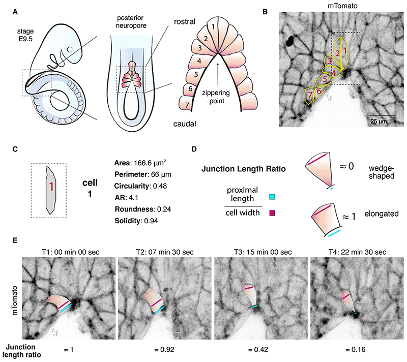
Zippering is a phenomenon of tissue morphogenesis whereby fusion between opposing epithelia progresses unidirectionally over significant distances, similar to the travel of a zip fastener, to ultimately ensure closure of an opening. A comparable process can be observed during Drosophila dorsal closure and mammalian wound healing, while zippering is employed by numerous organs such as the optic fissure, palatal shelves, tracheoesophageal foregut, and presumptive genitalia to mediate tissue sealing during normal embryonic development. Particularly striking is zippering propagation during neural tube morphogenesis, where the fusion point travels extensively along the embryonic axis to ensure closure of the neural tube. Advances in time-lapse microscopy and culture conditions have opened the opportunity for successful imaging of whole-mouse embryo development over time, providing insights into the precise cellular behavior underlying zippering propagation. Studies in mouse and the ascidian Ciona have revealed the fine-tuned cell shape changes and junction remodeling which occur at the site of zippering during neural tube morphogenesis. Here, we describe a step-by-step method for imaging at single-cell resolution the process of zippering and tissue remodeling which occurs during closure of the spinal neural tube in mouse. We also provide instructions and suggestions for quantitative morphometric analysis of cell behavior during zippering progression. This procedure can be further combined with genetic mutant models (e.g., knockouts), offering the possibility of studying the dynamics of tissue fusion and zippering propagation, which underlie a wide range of open neural tube defects.
拉链式组织形态发生是一种现象,即 opposing epithelia(上皮细胞)在相对的组织之间融合,并沿着长距离进行单向运动,类似于拉链的运动,最终确保开口的闭合。在果蝇背唇闭合和哺乳动物伤口愈合过程中可以观察到类似的过程,而在许多器官中,如视神经裂、腭板、气管食管前肠和假定的生殖器,拉链式组织形态发生用于介导组织密封,以确保正常胚胎发育过程中的组织完整性。在神经管形态发生过程中,拉链式组织形态发生的传播尤为引人注目,融合点沿着胚胎轴广泛传播,以确保神经管的闭合。延时显微镜和培养条件的进步为成功地对整个小鼠胚胎发育进行时间成像提供了机会,使我们深入了解了拉链式组织形态发生背后精确的细胞行为。在小鼠和海鞘 Ciona 中的研究揭示了在神经管形态发生过程中,在拉链式组织形态发生部位发生的精细的细胞形状变化和连接重塑。在这里,我们描述了一种一步一步的方法,用于以单细胞分辨率成像在小鼠脊柱神经管闭合过程中发生的拉链式组织形态发生和组织重塑过程。我们还提供了有关在拉链式组织形态发生过程中细胞行为的定量形态计量分析的说明和建议。此过程可以与遗传突变体模型(例如敲除)进一步结合,为研究组织融合和拉链式组织形态发生的动力学提供了可能性,这些动力学是广泛的神经管缺陷的基础。