Molecular Parasitology Laboratory, Queensland Institute of Medical Research, Queensland, Australia.
PLoS Negl Trop Dis. 2010 Feb 9;4(2):e598. doi: 10.1371/journal.pntd.0000598.
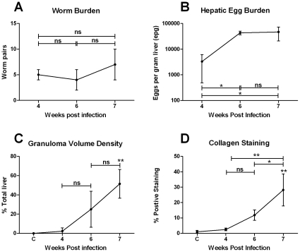
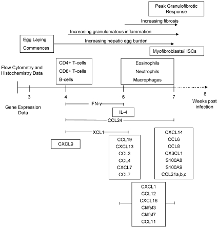
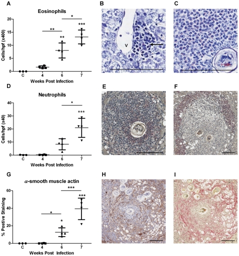
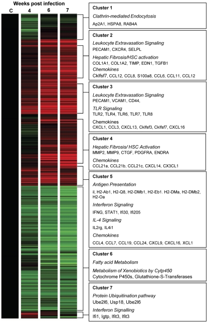
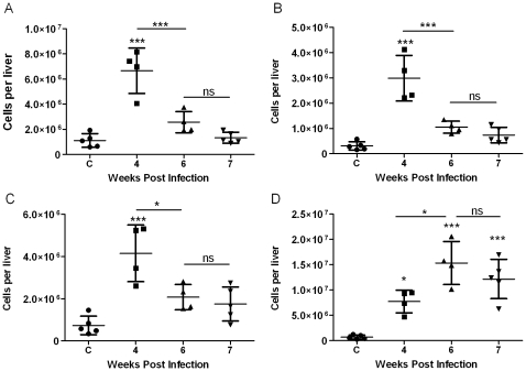
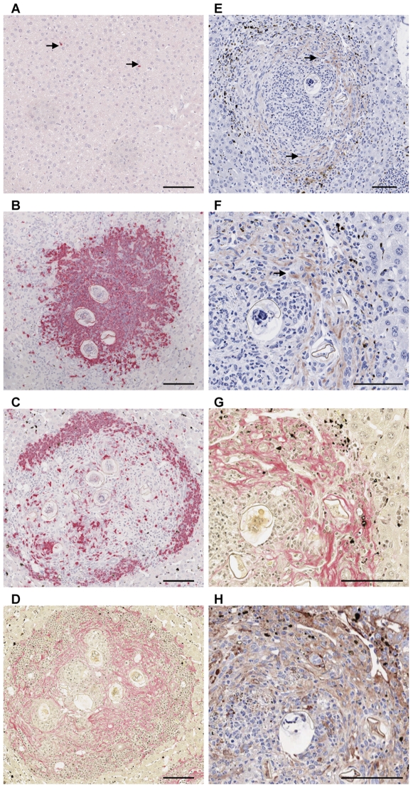
Schistosomiasis continues to be an important cause of parasitic morbidity and mortality world-wide. Determining the molecular mechanisms regulating the development of granulomas and fibrosis will be essential for understanding how schistosome antigens interact with the host environment. We report here the first whole genome microarray analysis of the murine liver during the progression of Schistosoma japonicum egg-induced granuloma formation and hepatic fibrosis. Our results reveal a distinct temporal relationship between the expression of chemokine subsets and the recruitment of cells to the infected liver. Genes up-regulated earlier in the response included T- and B-cell chemoattractants, reflecting the early recruitment of these cells illustrated by flow cytometry. The later phases of the response corresponded with peak recruitment of eosinophils, neutrophils, macrophages and myofibroblasts/hepatic stellate cells (HSCs) and the expression of chemokines with activity for these cells including CCL11 (eotaxin 1), members of the Monocyte-chemoattractant protein family (CCL7, CCL8, CCL12) and the Hepatic Stellate Cell/Fibrocyte chemoattractant CXCL1. Peak expression of macrophage chemoattractants (CCL6, CXCL14) and markers of alternatively activated macrophages (e.g. Retnla) during this later phase provides further evidence of a role for these cells in schistosome-induced pathology. Additionally, we demonstrate that CCL7 immunolocalises to the fibrotic zone of granulomas. Furthermore, striking up-regulation of neutrophil markers and the localisation of neutrophils and the neutrophil chemokine S100A8 to fibrotic areas suggest the involvement of neutrophils in S. japonicum-induced hepatic fibrosis. These results further our understanding of the immunopathogenic and, especially, chemokine signalling pathways that regulate the development of S. japonicum-induced granulomas and fibrosis and may provide correlative insight into the pathogenesis of other chronic inflammatory diseases of the liver where fibrosis is a common feature.
血吸虫病仍然是全球寄生虫发病率和死亡率的重要原因。确定调节肉芽肿和纤维化发展的分子机制对于了解血吸虫抗原如何与宿主环境相互作用至关重要。我们在此报告了日本血吸虫卵诱导的肉芽肿形成和肝纤维化过程中鼠肝的全基因组微阵列分析的首次研究。我们的结果揭示了趋化因子亚类的表达与细胞向感染肝脏募集之间的明显时间关系。在反应早期上调的基因包括 T 细胞和 B 细胞趋化因子,这反映了通过流式细胞术说明的这些细胞的早期募集。反应的后期阶段与嗜酸性粒细胞、中性粒细胞、巨噬细胞和肌成纤维细胞/肝星状细胞(HSCs)的最大募集以及对这些细胞具有活性的趋化因子的表达相对应,包括 CCL11(嗜酸性粒细胞趋化因子 1)、单核细胞趋化蛋白家族(CCL7、CCL8、CCL12)的成员和肝星状细胞/成纤维细胞趋化因子 CXCL1。在这个后期阶段,巨噬细胞趋化因子(CCL6、CXCL14)和替代激活的巨噬细胞标志物(例如 Retnla)的表达峰值进一步证明了这些细胞在血吸虫病引起的病理中的作用。此外,我们证明 CCL7 免疫定位在肉芽肿的纤维化区域。此外,中性粒细胞标志物的显著上调以及中性粒细胞和中性粒细胞趋化因子 S100A8 的局部定位到纤维化区域表明中性粒细胞参与了日本血吸虫诱导的肝纤维化。这些结果进一步了解了调节日本血吸虫诱导的肉芽肿和纤维化发展的免疫发病机制,特别是趋化因子信号通路,并可能为其他以纤维化为共同特征的慢性肝脏炎症性疾病的发病机制提供相关见解。