Department of Immunology, School of Medicine, Yangtze University, Jingzhou, China.
Clinical Molecular Immunology Center, School of Medicine, Yangtze University, Jingzhou, China.
Front Immunol. 2020 Feb 18;11:61. doi: 10.3389/fimmu.2020.00061. eCollection 2020.
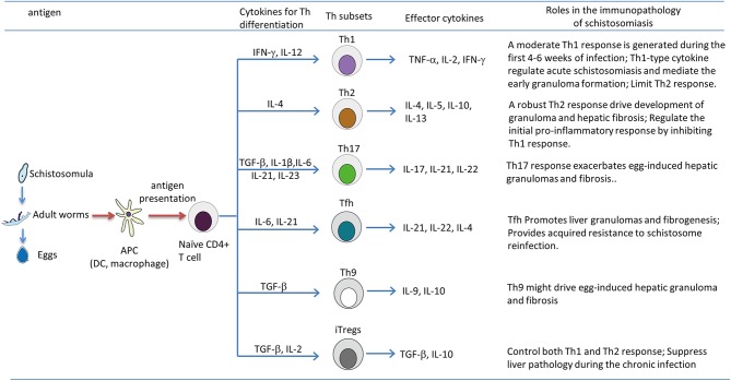
The parasitic worms, and , reside in the mesenteric veins, where they release eggs that induce a dramatic granulomatous response in the liver and intestines. Subsequently, infection may further develop into significant fibrosis and portal hypertension. Over the past several years, uncovering the mechanism of immunopathology in schistosomiasis has become a major research objective. It is known that T lymphocytes, especially CD4 T cells, are essential for immune responses against species. However, obtaining a clear understanding of how T lymphocytes regulate the pathological process is proving to be a daunting challenge. To date, CD4 T cell subsets have been classified into several distinct T helper (Th) phenotypes including Th1, Th2, Th17, T follicular helper cells (Tfh), Th9, and regulatory T cells (Tregs). In the case of schistosomiasis, the granulomatous inflammation and the chronic liver pathology are critically regulated by the Th1/Th2 responses. Animal studies suggest that there is a moderate Th1 response to parasite antigens during the acute stage, but then, egg-derived antigens induce a sustained and dominant Th2 response that mediates granuloma formation and liver fibrosis. In addition, the newly discovered Th17 cells also play a critical role in the hepatic immunopathology of schistosomiasis. Within the liver, Tregs are recruited to hepatic granulomas and exert an immunosuppressive role to limit the granulomatous inflammation and fibrosis. Moreover, recent studies have shown that Tfh and Th9 cells might also promote liver granulomas and fibrogenesis in the murine schistosomiasis. Thus, during infection, T-cell subsets undergo complicated cross-talk with antigen presenting cells that then defines their various roles in the local microenvironment for regulating the pathological progression of schistosomiasis. This current review summarizes a vast body of literature to elucidate the contribution of T lymphocytes and their associated cytokines in the immunopathology of schistosomiasis.
寄生虫蠕虫 和 ,存在于肠系膜静脉中,在那里它们释放卵,诱导肝脏和肠道中的剧烈肉芽肿反应。随后,感染可能进一步发展为显著的纤维化和门静脉高压。在过去的几年中,揭示血吸虫病免疫病理学的机制已成为一个主要的研究目标。已知 T 淋巴细胞,特别是 CD4 T 细胞,对于针对 物种的免疫反应至关重要。然而,要清楚地了解 T 淋巴细胞如何调节病理过程,这被证明是一个艰巨的挑战。迄今为止,CD4 T 细胞亚群已被分为几种不同的 T 辅助(Th)表型,包括 Th1、Th2、Th17、滤泡辅助 T 细胞(Tfh)、Th9 和调节性 T 细胞(Treg)。在血吸虫病中,肉芽肿炎症和慢性肝病理由 Th1/Th2 反应严格调节。动物研究表明,在急性阶段,寄生虫抗原会引起适度的 Th1 反应,但随后,卵衍生的抗原会诱导持续和占主导地位的 Th2 反应,介导肉芽肿形成和肝纤维化。此外,新发现的 Th17 细胞也在血吸虫病的肝免疫病理学中发挥关键作用。在肝脏中,Treg 被招募到肝肉芽肿中,并发挥免疫抑制作用,以限制肉芽肿炎症和纤维化。此外,最近的研究表明,Tfh 和 Th9 细胞也可能促进小鼠血吸虫病中的肝肉芽肿和纤维发生。因此,在感染过程中,T 细胞亚群与抗原呈递细胞发生复杂的相互作用,从而确定它们在局部微环境中的各种作用,以调节血吸虫病的病理进展。本综述总结了大量文献,阐明了 T 淋巴细胞及其相关细胞因子在血吸虫病免疫病理学中的作用。