Department of Anesthesiology, School of Medicine and Public Health, University of Wisconsin, 1300 University Avenue, Madison, WI 53706, USA.
Pain. 2010 Apr;149(1):152-159. doi: 10.1016/j.pain.2010.02.001. Epub 2010 Feb 18.
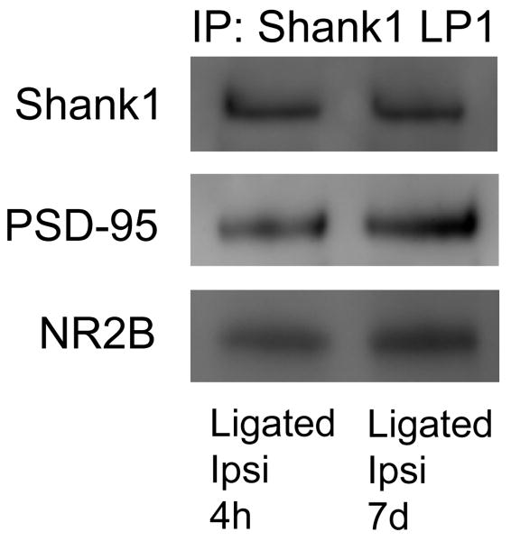
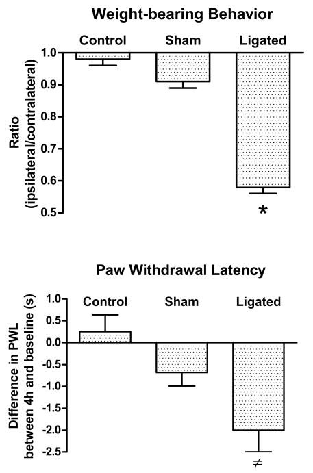
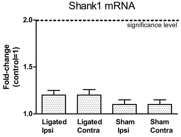
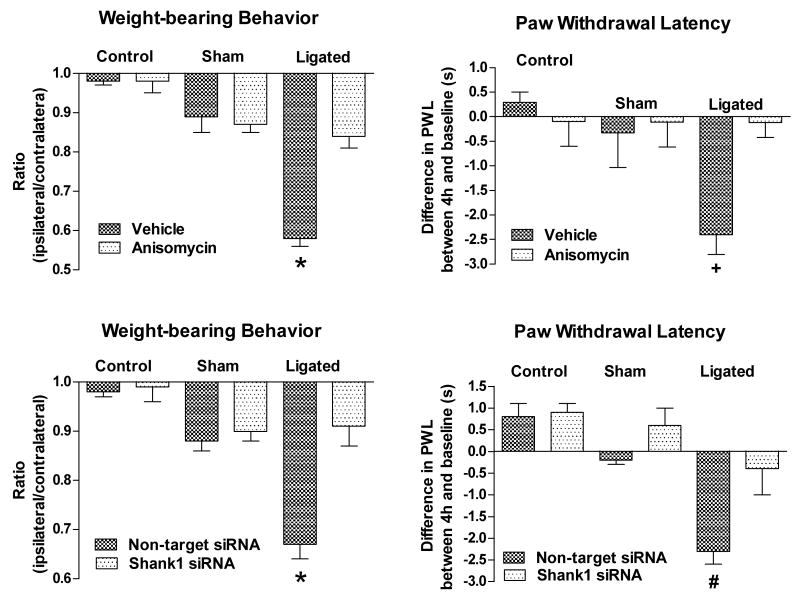
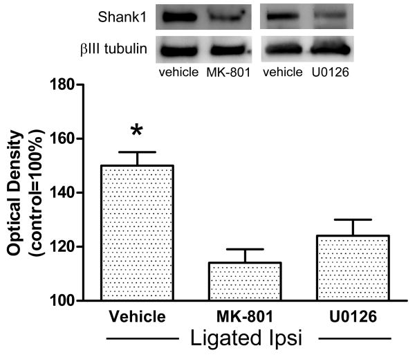
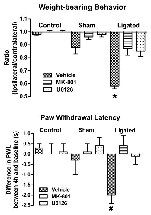
Plasticity in the spinal dorsal horn may contribute to the development of pain following peripheral nerve injury. Shank proteins are a constituent family of the post-synaptic density (PSD), and they may play a role in synaptic plasticity through activity-dependent synaptic remodeling and growth. In this study we examined the early consequences of the loose ligation of the sciatic nerve on Shank1 protein and message levels in the PSD of spinal dorsal horn neurons. Four hours after sciatic ligation, the protein levels of Shank1 increased in the ipsilateral PSD of ligated animals. In contrast, no changes were detected in the contralateral PSD of these ligated animals, or either the ipsilateral or contralateral PSD of sham-operated animals. Shank1 was linked to the PSD marker protein PSD-95 and the NR2B subunit of NMDA receptors. The ligated animals also exhibited two early signs of pain behavior, a shift in weight distribution and thermal hyperalgesia. There was no overall change in Shank1 message in either ligated or sham-operated animals. The accumulation of Shank1 in the PSD was abolished by intrathecal pre-treatment with anisomycin or Shank1 siRNA, but not with non-target siRNA. The same pre-treatment prevented both the early signs of pain behavior. Intrathecal pre-treatment with either MK-801 or U0126 similarly prevented the Shank1 accumulation and alleviated both the behavioral signs of pain. The early accumulation of Shank1 in the PSD of dorsal horn neurons may be a necessary step in the injury-associated plasticity that in time leads to the development of persistent pain.
脊髓背角的可塑性可能有助于外周神经损伤后疼痛的发展。Shank 蛋白是突触后密度(PSD)的组成家族之一,它们可能通过活性依赖性突触重塑和生长在突触可塑性中发挥作用。在这项研究中,我们研究了坐骨神经松解对脊髓背角神经元 PSD 中 Shank1 蛋白和 mRNA 水平的早期影响。坐骨神经结扎后 4 小时,结扎动物同侧 PSD 中的 Shank1 蛋白水平增加。相比之下,在这些结扎动物的对侧 PSD 中或假手术动物的同侧或对侧 PSD 中均未检测到变化。Shank1 与 PSD 标记蛋白 PSD-95 和 NMDA 受体的 NR2B 亚基相连。结扎动物还表现出两种疼痛行为的早期迹象,即体重分布的改变和热痛觉过敏。无论是结扎动物还是假手术动物,Shank1 的 mRNA 都没有总体变化。鞘内预先用放线菌酮或 Shank1 siRNA 处理可消除 PSD 中 Shank1 的积累,但非靶向 siRNA 则不能。相同的预处理可预防疼痛行为的早期迹象。鞘内预先用 MK-801 或 U0126 处理也可防止 Shank1 积累,并缓解疼痛的行为迹象。背角神经元 PSD 中 Shank1 的早期积累可能是与损伤相关的可塑性的必要步骤,这种可塑性最终导致持续性疼痛的发展。