Cell Isolation and Transplantation Center, Department of Surgery, Geneva University Hospitals and University of Geneva, Geneva, Switzerland.
Diabetes. 2010 May;59(5):1202-10. doi: 10.2337/db09-1177. Epub 2010 Feb 25.
It is generally admitted that the endocrine cell organization in human islets is different from that of rodent islets. However, a clear description of human islet architecture has not yet been reported. The aim of this work was to describe our observations on the arrangement of human islet cells.
Human pancreas specimens and isolated islets were processed for histology. Sections were analyzed by fluorescence microscopy after immunostaining for islet hormones and endothelial cells.
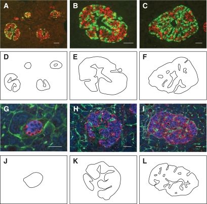
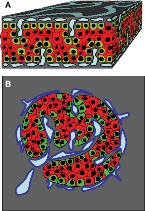
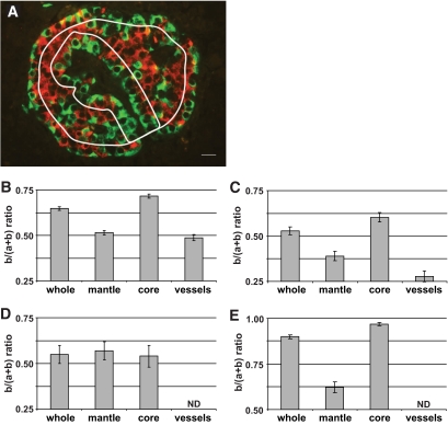
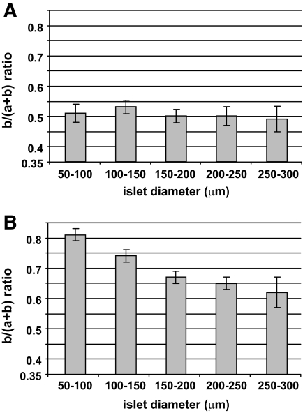
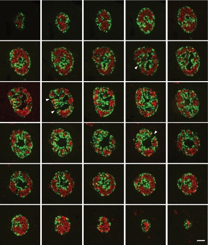
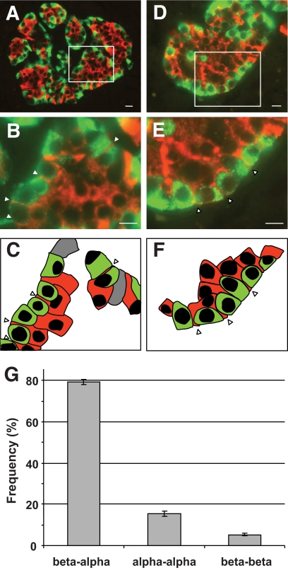
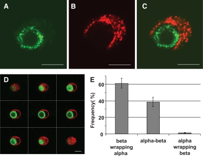
In small human islets (40-60 mum in diameter), beta-cells had a core position, alpha-cells had a mantle position, and vessels laid at their periphery. In bigger islets, alpha-cells had a similar mantle position but were found also along vessels that penetrate and branch inside the islets. As a consequence of this organization, the ratio of beta-cells to alpha-cells was constantly higher in the core than in the mantle part of the islets, and decreased with increasing islet diameter. This core-mantle segregation of islet cells was also observed in type 2 diabetic donors but not in cultured isolated islets. Three-dimensional analysis revealed that islet cells were in fact organized into trilaminar epithelial plates, folded with different degrees of complexity and bordered by vessels on both sides. In epithelial plates, most beta-cells were located in a central position but frequently showed cytoplasmic extensions between outlying non-beta-cells.
Human islets have a unique architecture allowing all endocrine cells to be adjacent to blood vessels and favoring heterologous contacts between beta- and alpha-cells, while permitting homologous contacts between beta-cells.
人们普遍承认,人类胰岛中的内分泌细胞组织与啮齿动物胰岛中的不同。然而,尚未有关于人胰岛结构的明确描述。本研究旨在描述我们对人胰岛细胞排列的观察结果。
对人胰腺标本和分离的胰岛进行组织学处理。免疫染色胰岛激素和内皮细胞后,通过荧光显微镜分析切片。
在较小的人胰岛(直径 40-60 微米)中,β细胞位于核心位置,α细胞位于外套位置,血管位于其周围。在较大的胰岛中,α细胞具有相似的外套位置,但也存在于穿透和分支进入胰岛内部的血管周围。由于这种组织排列,β细胞与α细胞的比例在胰岛的核心部分始终高于外套部分,并且随着胰岛直径的增加而降低。这种胰岛细胞的核心-外套分离在 2 型糖尿病供体中也观察到,但在培养的分离胰岛中未观察到。三维分析显示,胰岛细胞实际上被组织成三层上皮板,以不同程度的复杂性折叠,并在两侧由血管包围。在上皮板中,大多数β细胞位于中央位置,但经常在外围非β细胞之间出现细胞质延伸。
人胰岛具有独特的结构,允许所有内分泌细胞与血管相邻,并有利于β-和α-细胞之间的异源接触,同时允许β-细胞之间的同源接触。